Researchers have built a computer-aided detection (CAD) algorithm that detects bladder cancer lesions with high sensitivity and specificity by analyzing 3D MRI images. They have unveiled the full details in the International Journal of Computer Assisted Radiology and Surgery.
The system uses MR virtual cystoscopy images to detect and extract 3D regions of bladder tumors, according to lead author Dan Xiao and colleagues. The algorithm uses a novel method of measuring wall thickness that contributes to higher sensitivity and fewer false positives, according to the study team from the Fourth Military Medical University and Tangdu Hospital in Shaanxi, China, and City University of New York/College of Staten Island in New York City.
"The experimental result demonstrates the feasibility of the proposed pipeline on the detection and extraction of bladder tumors," they wrote. "It may provide an effective way to achieve the goal of evaluating the whole bladder for tumor detection and local staging."
Techniques need improvement
Bladder carcinoma is not only the most common cancer of the urinary system; it is the fourth most common cancer and the eighth leading cause of death among men. And it recurs frequently, wrote Xiao and colleagues (IJCARS, 20 June 2015).
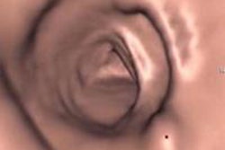
The gold standard for evaluating tumors, optical cystoscopy (OC), unfortunately can cause discomfort and even iatrogenic injuries. As a result, virtual cystography using CT or MR has recently become a promising alternative for noninvasively evaluating the bladder wall and urethral orifice as it "mimics the structure of true bladder to enable urologists inspect the whole bladder directly," Xiao et al wrote. In fact, studies have shown no differences in the results of conventional cystography versus the virtual technique for tumors 5 mm and larger.
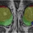
The group's process begins with the simultaneous segmentation of both inside and outside surfaces of 3D bladder volumes acquired at MRI. To detect bladder abnormalities, the scheme applies four volume-based morphological features to the MR volume -- and a novel fifth one designed to reflect differences between the inner and outer wall -- that together signal the presence of bladder tumors. In 10 patients, sensitivity was 100% with high specificity, the authors wrote.
CAD based on image features can help radiologists improve accuracy and efficiency. Previous studies used morphological features of the bladder such as curvedness, wall thickness, and bent rate to boost the accuracy of tumor detection, or more recently, bladder wall-thickness mapping, the team wrote.
But challenges have arisen in detecting tumors without too many false positives. And it has been difficult to capture whole tumor regions precisely from the bladder wall, essential for local staging and for predicting wall and perivesical infiltration, they added.
Three steps
In the current paper, the group sought to address those problems with a proposed CAD algorithm that performs bladder wall segmentation, seed region detection, and tumor region extraction. In step 1, the coupled directional level-set method is used to segment the inner and outer surfaces of bladder wall simultaneously from T2-weighted MR images.
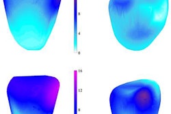
Step 2 calculates bladder wall thickness (BWT), curvedness (CV), shape index (SI), and bent rate (BR), the latter being a description of the geometric properties of a surface traditionally used to protect protruding objects. These values are compared with filtering criteria to determine whether a voxel is a seed or not, and each area composed of connecting seeds forms a seed region on the inner surface.
Step 3 uses detected seeds in a region as starting points to form a 3D candidate region using all wall points on the tracing. Then fuzzy c-means clustering with spatial information (sFCM) is used to extract whole tumor regions from the surrounding bladder wall, Xiao et al wrote.
A novel morphological feature defined by the bent rate difference (BRD) of corresponding wall voxels on the inner and outer surfaces of the bladder wall has shown good performance in detecting locally thickened regions, the authors wrote. Combined with previously used morphological features, this feature can be used for candidate detection, improving sensitivity and reducing false-positives. Finally, fuzzy c-means segmentation with spatial information is used to further extract tumors from the surrounding wall tissues.
"For a small bump protruding out of the bladder wall, the measurement of its thickness as the distance between the two surfaces tends to be a good indicator of the occurrence of abnormalities," the authors wrote. "Traditional distance metrics, such as
Hausdorff distance and distance transform model may fail in determining unique and nonintersecting paths between surface regions with protruded lesions."
The CAD technique was tested on T2-weighted MRI datasets of 10 patients with bladder cancer. Images were acquired on a Discovery MR750 (GE Healthcare) scanner equipped with a phased-array body coil, with an axial 3D-cube T2 sequence, chosen for its good tissue contrast and relatively fast acquisition. Other parameters included 512 x 512 matrix size, 0.5-mm pixel spacing, 1-mm slice thickness, TE = 135 ms, and TR = 2,500 ms. Patients were instructed to drink enough water to fill the bladder before imaging.
The datasets contained 16 tumors ranging in size from 5 mm to 33 mm. Three tumors were superficial while others had invaded the bladder wall. Experienced radiologists delineated each tumor manually slice-by-slice, which was used as the reference standard for the CAD.
In order to determine an optimal feature combination for tumor detection, the study team evaluated the performance of combinations of the four features, evaluating them with different filtering criteria and combining them with the bent rate difference, which measures the curving similarity of the inner and outer bladder wall surfaces.
Perfect sensitivity, some false-positives
The authors found that preliminary evaluation using volumetric MR bladder images of patients validated its superior performance in sensitivity improvement and false-positive reduction, with 100% sensitivity and 2.3 false positives per case, the authors wrote.
Compared with tumor regions traced by expert radiologists, the overlap rate for tumors delineated by the CAD was 86.3%, the study team reported.
For accurate extraction of tumor tissues, fuzzy c-means clustering with spatial information was used to further extract tumors from surrounding wall tissues in candidate regions. The experimental result demonstrated the feasibility of the CAD algorithm on the detection and extraction of bladder tumors.
Detection of early-stage bladder abnormalities remains a challenge with current MRI scanners, but the proposed CAD pipeline for MR virtual cystography has shown potential for evaluating the entire bladder wall for detecting tumors and managing recurrence, the authors wrote. With its ability to accurately delineate tumor regions, the method may also be used for local staging of bladder cancer. But additional research is needed to prove its effectiveness over time.
"More evaluation should be performed to validate the performance and robustness of the proposed pipeline on larger clinical datasets," Xiao et al wrote. "It is expected that the performance could be further improved with the improvement on MRI spatial resolution and contrast between the tumor and its surrounding wall tissues."
