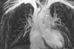
Visualizing CT images of their own coronary artery calcifications prompts patients to make the important lifestyle changes they need to stay healthy, according to a new Danish study presented this week at the EuroHeartCare meeting in Dubrovnik, Croatia.
The study looked at 189 patients with hyperlipidemia who underwent CT coronary angiography (CCTA) and were diagnosed with nonobstructive coronary artery disease. Half were randomized to an intervention group that was shown the calcifications in a counseling session aimed at education and lifestyle modification. The other half received standard follow-up. The counseled patients were significantly more likely to change their lifestyles.
"Seeing their calcified coronary arteries on the CT image was clearly an eye-opener for patients," noted study presenter Rikke Elmose Mols, a nurse and PhD student at Aarhus University Hospital, in a statement. "We received comments such as, 'It is my coronary artery and my coronary artery calcification and I am facing a real risk and challenge.' This may be the wake-up call patients need to take their medication and modify their behaviors to reduce their risk of having a coronary artery event."
Images of calcified arteries like these can inspire people to change. LMC = left main coronary artery. Ao = aorta. LAD = left anterior descending coronary artery. Image courtesy of Rikke Elmose Mols.The study aimed to evaluate the influence of visualization of coronary artery calcifications along with standard information about risk and lifestyle modification about serum cholesterol levels and other risk factors in patients with hyperlipidemia who had just been diagnosed with nonobstructive coronary artery disease.
The intervention group received a 25-minute consultation with a nurse focusing on communication of risk and lifestyle changes, with patients seeing images of their own calcifications on CT. The counseling session included 15 minutes with a nurse explaining the association between coronary artery calcification and an increased risk of future cardiovascular events, as well as the association between cardiovascular risk factors and the development of coronary artery calcifications. The last 10 minutes were devoted to advice about statins, aspirin, blood pressure, a healthy diet, exercise, and quitting smoking, all based on European Society of Cardiology (ESC) prevention guidelines.
Forty-four patients who discontinued statin therapy due to side effects were excluded from analysis at a risk-factor follow-up session six months after the imaging. Results in the remaining patients revealed:
- Significantly greater reductions of plasma cholesterol in the intervention group (1.71 mmol/L) compared with controls (1.44 mmol/L, p = 0.027).
- More patients continued smoking in the control group (22%) compared with the intervention group (9%, p = 0.014).
- More control group patients (64%) continued eating an unhealthy diet versus those in the intervention group (44%, p = 0.005).
- An average 0.5 kg weight gain in the control group versus an average 1.5 kg weight loss in the intervention group.
- A trend toward greater adherence to statin therapy in the intervention group (p = 0.056).
Visualizing their health threat motivates patients to make changes to reduce their risk, the study team concluded. Patients who saw images of their calcified coronary arteries were more likely to stop smoking, lose weight, eat a healthy diet, take recommended statins, and reduce their plasma cholesterol levels.
A larger study is needed to confirm the findings and evaluate the cost-effectiveness of implementing postscan counseling in clinical practice, they added.


![Examples of ultrasound findings and techniques. (A) Images in a 39-year-old male patient with a mass in the left thigh. The mass is heterogeneous on the B-mode US image (compared with the patient in D) and showed increased microvascularity (superb microvascular imaging [SMI]) and shear-wave elastography (SWE) values. Undifferentiated pleomorphic sarcoma was diagnosed at biopsy (with pleomorphic rhabdomyosarcoma in surgical specimen). (B) Images in an 18-year-old male patient with a mass in the left leg. The mass is hypoechoic on the B-mode image, with no other findings suggestive of malignancy. The lesion is in contact with the cortex of the tibia, which is slightly irregular. CT revealed a doubtful anteromedial tibial erosion. The microvascular study demonstrated high vascularization, suggestive of malignancy. Periosteal Ewing sarcoma was diagnosed with both histologic and immunohistochemical confirmation. (C) Images in a 69-year-old female patient with a lump growing on the outside of the left leg. Multiple SWE examinations were performed (please note the high values obtained in the measurements, whereas the color map highlights the stiffness relative to adjacent tissues). SMI showed areas of increased vascularization to target for sampling. Undifferentiated spindle cell sarcoma was diagnosed at biopsy, with residual leiomyosarcoma in the surgical specimen after neoadjuvant therapy. (D) Images in a 56-year-old female patient with a mass in the right thigh. The mass is heterogeneous at both B-mode ultrasound (similar to patient A) and MRI (coronal T2-weighted spectral attenuated inversion recovery [SPAIR]; T1-weighted pre-contrast and postcontrast imaging), which even shows uptake after the administration of paramagnetic contrast material, which is traditionally suggestive of malignancy. Low values at SMI and elastography are suggestive of benignity. Spindle cell lipoma was diagnosed at biopsy, with atypical spindle cell lipomatous tumor in the surgical specimen.](https://img.auntminnieeurope.com/mindful/smg/workspaces/default/uploads/2026/08/images-radiol250278fig2.APCFLSvX6p.jpg?auto=format%2Ccompress&fit=crop&h=100&q=70&w=100)


![Examples of ultrasound findings and techniques. (A) Images in a 39-year-old male patient with a mass in the left thigh. The mass is heterogeneous on the B-mode US image (compared with the patient in D) and showed increased microvascularity (superb microvascular imaging [SMI]) and shear-wave elastography (SWE) values. Undifferentiated pleomorphic sarcoma was diagnosed at biopsy (with pleomorphic rhabdomyosarcoma in surgical specimen). (B) Images in an 18-year-old male patient with a mass in the left leg. The mass is hypoechoic on the B-mode image, with no other findings suggestive of malignancy. The lesion is in contact with the cortex of the tibia, which is slightly irregular. CT revealed a doubtful anteromedial tibial erosion. The microvascular study demonstrated high vascularization, suggestive of malignancy. Periosteal Ewing sarcoma was diagnosed with both histologic and immunohistochemical confirmation. (C) Images in a 69-year-old female patient with a lump growing on the outside of the left leg. Multiple SWE examinations were performed (please note the high values obtained in the measurements, whereas the color map highlights the stiffness relative to adjacent tissues). SMI showed areas of increased vascularization to target for sampling. Undifferentiated spindle cell sarcoma was diagnosed at biopsy, with residual leiomyosarcoma in the surgical specimen after neoadjuvant therapy. (D) Images in a 56-year-old female patient with a mass in the right thigh. The mass is heterogeneous at both B-mode ultrasound (similar to patient A) and MRI (coronal T2-weighted spectral attenuated inversion recovery [SPAIR]; T1-weighted pre-contrast and postcontrast imaging), which even shows uptake after the administration of paramagnetic contrast material, which is traditionally suggestive of malignancy. Low values at SMI and elastography are suggestive of benignity. Spindle cell lipoma was diagnosed at biopsy, with atypical spindle cell lipomatous tumor in the surgical specimen.](https://img.auntminnieeurope.com/mindful/smg/workspaces/default/uploads/2026/08/images-radiol250278fig2.APCFLSvX6p.jpg?auto=format%2Ccompress&fit=crop&h=112&q=70&w=112)