The changing role of our modern imaging techniques can be really interesting. Traditionally our radiological techniques were used to confirm a clinical diagnosis, and because these techniques were often quite invasive, they were only applied when there was a reasonable chance of the examination being positive.
 Figure 1: Right-sided opacity is shown separately from the ureter, proving it is a phlebolith.
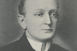
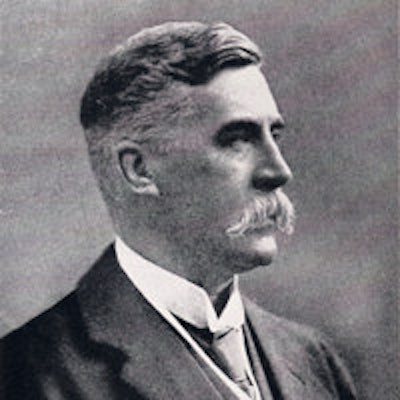
In the diagnosis of ureteric calculi, the technique in the late 19th century as used by Dr. Edwin Hurry Fenwick (1856-1944), the British urologist and pioneer of cystoscopy, is worth recalling.1 Prior to the use of radiography, the surgeon performed a cystoscopy and a ureteric bougie with wax on its tip was passed up the ureter. If the wax was scratched when removed, this was evidence of the presence of a stone. This was not a technique to be undertaken lightly, nor was it particularly accurate.
Following the discovery of the x-ray in 1895, Hurry Fenwick was an enthusiastic supporter of the new technique.2 A radiopaque ureteric bougie was passed and a radiograph was made. The position of an opacity in relation to the ureter could be determined with confidence and a phlebolith distinguished from a ureteric calculus (figure 1).
He commented on the distressing situation with the failure of operative surgery when a kidney was opened and damaged to remove a stone when it was no longer in the kidney and was now in the ureter. He estimated that this happened in about 30% of cases when the "x-ray expert" was not called upon to help in the diagnosis. The "x-ray expert" (radiologist) can "guide the urinary surgeon [urologist] with a precision unattainable before the introduction of the [x-ray] method is without cavil."
Hurry Fenwick was writing in 1908 when the techniques used were still quite primitive and before the introduction of retrograde or intravenous pyelography.
 Figure 2, above: Blackboard diagram of radiographic findings. Figure 3 (below): Cystoscopic phantom from the electro-medical instruments document of K Schall (1899).
He was a remarkable surgeon and was a pioneer of cystoscopy and also of the use of radiography for urological abnormalities. He was one of the first to practice clinical-radiological-pathological correlation (figure 2), correlating the clinical findings with the radiography and then with the operative findings. Quite remarkably, he was teaching operative cystoscopy using a bladder phantom before 1900 (figure 3).
One of the problems of modern radiology is that with the increasing availability of techniques, the level of indication for a given procedure has dropped. So a CT scan of the head is now used as part of a confusion screen, and even a short time ago a CT head scan was difficult to request without specific indications.
 Figure 4: Coronal CT shows a left ureteric calculus.
A traditional invasive technique such as a lumbar myelogram or radiculogram would only be used if the likelihood of finding an abnormality was quite high. Part of the problem is that as the threshold for investigation falls, the significance of radiological findings is more difficult to determine. So with a high clinical probability of an abnormality, the utility of MRI scanning in the lumbar spine is good and there is a high chance of finding abnormalities that may be surgically treated. As scanning is now used for more general causes of back pain, the clinical significance of the findings are more difficult to determine.
Figure 4 is a CT scan performed for a clinical diagnosis of complicated diverticular disease. The coronal CT scan showed the left-sided ureteric calculus with hydronephrosis. Within a couple of hours, the patient was having a nephrostomy for an obstructed septic kidney. Whilst Hurry Fenwick would be astonished by our ability to perform a coronal CT to show the ureteric calculus and then to drain a compromised septic kidney, I think he would be concerned about the clinical confusion of diverticular disease and renal disease. Radiology works best following accurate clinical assessment. The role of radiology is to confirm and support a clinical diagnosis, and not to rule out less-likely diagnoses by an indiscriminate use of complex radiological examinations.
Editor's note: Portrait photo on home page is courtesy of the British Association of Urological Surgeons.
Dr. Adrian Thomas is chairman of the International Society for the History of Radiology and honorary librarian at the British Institute of Radiology.
References
- Hurry Fenwick E. The Cardinal Symptoms of Urinary Disease: Their Diagnostic Significance and Treatment. London, U.K.: J&A Churchill; 1893.
- Hurry Fenwick E. The Value of Radiography in the Diagnosis and Treatment of Urinary stone: A Study in Clinical and Operative Surgery. London, U.K.: J&A Churchill; 1908.
The comments and observations expressed herein do not necessarily reflect the opinions of AuntMinnieEurope.com, nor should they be construed as an endorsement or admonishment of any particular vendor, analyst, industry consultant, or consulting group.