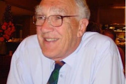
With the passing of Dr. Ronald Grainger (1922-2014) in the autumn, radiology lost one of its greats. He was joint editor of the profession's "bible," Grainger & Allison's Diagnostic Radiology, and the first professor of radiology in Sheffield, U.K. 1
 Dr. Ronald Grainger (1922-2014).
Grainger qualified in medicine at Leeds in 1945. He initially worked in medicine, but on meeting the late Robert Steiner, whom he called "the master radiologist of his generation," his interest in radiology was kindled.
Steiner was working in Sheffield at that time, and was giving tutorials that Grainger attended. He started his radiology training in 1952 in Sheffield, subsequently moving to London to work with Sir Peter Kerley (of "B lines" fame). He returned to Sheffield and was appointed consultant radiologist on 1 October 1960. In Sheffield, he developed an interest in cardiovascular radiology and intravascular contrast media, and became an internationally recognized expert in both areas.
In 1962, Grainger founded a radiological club, the 62 Club. This club was, and remains, very successful and celebrated its 50th anniversary with a meeting in Newquay, Cornwall, in 2012.
 The 62 Club celebrated its 50th anniversary in 2012.
Grainger had been a co-editor of the first edition of the well-known textbook of radiology edited by David Sutton. Grainger subsequently produced his own textbook of radiology and chose Dr. David Allison, from London's Hammersmith Hospital, as his co-editor. The book was first published in 1986, and Grainger was to see the appearance of the sixth edition, published in 2014.
The authors of "Grainger and Allison" -- known simply as G&A -- were drawn from the members of the 62 Club. G&A has now introduced many generations to the world of radiology, and like numerous radiologists, I have the latest edition on my Kindle!
Grainger developed a deep interest in the history of the development of intravascular contrast media, and this was demonstrated in his masterly Mackenzie Davidson Memorial Lecture, which he gave to the British Institute of Radiology (BIR) in 1981.2 In the lecture, he celebrated those who developed modern contrast media, and described current advances.
Grainger was very keen to give due credit to the work of Moses Swick, whose contributions had been somewhat neglected, he thought. The paper is well worth reading even today. It was a personal pleasure and privilege for me to write a book chapter on the development of contrast media with Grainger, and he was a courteous and helpful co-author.3
 Grainger co-edited a book about pioneers in angiography, and worked closely with the Portuguese School of Angiography.
I was introduced to the Portuguese School of Angiography in the lovely little book that Grainger edited with the Portuguese anglophile, Dr. J.A. Viega-Pires.4 The period of the late 1920s in Portugal was a very fruitful period for radiology, with famous names such as Egas Moniz and Reynaldo dos Santos. Many techniques were developed, such as cerebral angiography and lumbar aortography. This volume, which ran to a second edition, is one of my favorite books on the history of radiology.
Ronald Grainger was a charming man and he will be deeply missed by many people.
Dr. Adrian Thomas is chairman of the International Society for the History of Radiology and honorary librarian at the British Institute of Radiology.
References
- Allison D., Grainger RG. Newsletter of the Royal College of Radiologists. Winter 2014;(113):15.
- Grainger RG. Intravascular contrast media -- the past, the present, and the future. Br J Radiol. 1982;55(649):1-18.
- Grainger RG, Thomas AMK. History of the development of radiological contrast media (1895-1996). In Dawson P, Cosgrove DO, Grainger RG, eds. Textbook of Contrast Media. Oxford: Isis; 1999:3-14.
- Viega-Pires JA, Grainger RG. Pioneers in Angiography: The Portuguese School of Angiography. Lancaster: Isis;1999.
The comments and observations expressed herein do not necessarily reflect the opinions of AuntMinnieEurope.com, nor should they be construed as an endorsement or admonishment of any particular vendor, analyst, industry consultant, or consulting group.