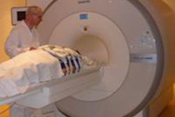
Using functional MRI (fMRI), researchers found that pornography triggered brain activity in people with compulsive sexual behavior similar to the brain activity seen in drug addicts when they are triggered by drugs.
The group from the University of Cambridge and other institutions imaged brain activity in 19 male patients with compulsive sexual behavior and compared them to the same number of healthy volunteers. The patients started watching pornography at earlier ages and in higher proportions relative to the healthy volunteers. The patients also had substantial difficulties controlling their sexual behavior, which affected their lives and relationships (PLOS One, July 11, 2014).
The study participants were shown a series of short videos featuring either sexually explicit content or sports while their brain activity was monitored using fMRI.
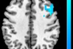
Lead author Dr. Valerie Voon and colleagues found that three regions -- the ventral striatum, dorsal anterior cingulate, and amygdala -- were more active in the brains of the people with compulsive sexual behavior than the healthy volunteers. These regions are also particularly activated in drug addicts when they are shown drug stimuli.
The ventral striatum is involved in processing reward and motivation, while the dorsal anterior cingulate is implicated in anticipating rewards and drug craving. The amygdala is involved in processing the significance of events and emotions.
The researchers cautioned that the findings do not necessarily mean that pornography itself is addictive. Much more research is needed to understand the relationship between compulsive sexual behavior and drug addiction, Voon noted.