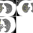
CT screening of high-risk individuals for lung cancer can identify malignant nodules sooner, but every CT scan performed, even at a low dose, confers a small risk of causing cancer as a result of cumulative radiation dose exposure. German researchers think MRI provides a viable alternative.
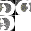
A multi-institutional team conducted a study to investigate the potential of MRI for detecting cancer-suspicious lung lesions. Dr. Gregor Sommer, from the department of radiology at the German Cancer Research Center (DKFZ) in Heidelberg, and coresearchers prospectively recruited 49 participants of the ongoing German Lung Cancer Screening and Intervention trial (LUSI) between January 2009 and July 2012. These 31 men and 18 women had either a pulmonary lesion larger than 10 mm or a lesion with calculated volume doubling time less than 400 days at follow-up. All had been immediately recalled for another CT exam according to LUSI protocol and, once enrolled in the study, an MRI exam (European Journal of Radiology, March 2014, Vol. 83:3, pp. 600-605).
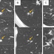
Thirty chest MRI datasets, recorded using identical sequences to those used for the study participants, were selected from healthy individuals. Two board-certified radiologists then interpreted the anonymized combined exams. The radiologists indicated the presence of suspicious lesions and their level of diagnostic confidence. For positive findings, they provided the size and position of each detected lesion and indicated the sequence that was most useful for diagnosis. A third radiologist unblinded the data and compared the positions of the MR-detected pulmonary nodules with the corresponding CT datasets.
The CT exams showed one suspicious lesion (with an average maximum diameter of 15 mm) in 46 studies, and two lesions in the remaining four datasets. On MRI, one radiologist identified half of these (27) lesions; the other radiologist identified 25. Two malignant nodules were missed by both radiologists. The false-negative ratio was 52%. The overall sensitivity of MRI was 48% (26/54) and the specificity was 88% (29/33). The sensitivity of the MR images was 78% for malignant nodules and 36% for benign ones.
Based on the findings of their study, the researchers believe that MRI has the potential to replace low-dose CT as the primary tool for lung cancer screening, provided that its sensitivity and specificity for detection of malignant lung nodules can be confirmed in further clinical trials as being greater than 75% and 90%, respectively. They pointed out that MRI offers the possibility of shortening screening intervals for individuals at very high risk. They also suggest that MRI may be used to supplement a low-dose CT study when clinical scenarios merit, due to its sensitivity for identifying malignant nodules.
"The expense of an MRI exam might avoid futile surgery or bronchoscopy by increasing the specificity and predictive value of the screening test," Sommer noted. "As these invasive procedures are much more expensive than any imaging exam, I see a potential benefit of using MRI instead of CT from an economic point of view."
The authors also suggested that simultaneous evaluation of MRI along with the large prospective CT screening trials to assess the benefit of cancer-specific mortality is warranted. However, Sommer does not anticipate this will happen soon. "As the prevalence of malignancy even in a high-risk population is very low, a prospective validation of MRI for lung cancer screening would require inclusion of thousands of individuals. This would require a huge logistical effort that could only be handled in a multicenter environment and would be very expensive," he said.
© IOP Publishing Limited. Republished with permission from medicalphysicsweb, a community website covering fundamental research and emerging technologies in medical imaging and radiation therapy.


