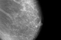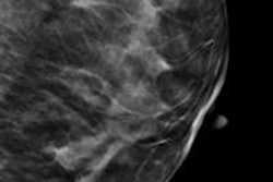
Synthetically reconstructed 2D images with digital breast tomosynthesis (DBT) was comparable to conventional 2D mammography with DBT for breast cancer screening, in a Norwegian study presented at this month's RSNA meeting in Chicago.
Taking full-field digital mammography (FFDM) images as well as tomosynthesis images doubles a woman's radiation dose, so using synthetic 2D images with tomosynthesis would be better for patients, the researchers noted.
Dr. Per Skaane, PhD, from the University of Oslo, and colleagues compared the diagnostic performance of full-field digital mammography (FFDM) plus DBT with that of synthesized 2D images plus DBT for breast cancer screening. The study is part of the Oslo Tomosynthesis Screening Trial, which began in 2010 and receives equipment and support from Hologic.
Skaane's team used Hologic's C-View 2D imaging software and Selenia Dimensions 3D breast tomosynthesis system for their research. The software is designed to enable tomosynthesis to produce 2D mammography images without the higher radiation dose typically involved in combined 2D/3D studies.
Are 2D images really necessary? Yes, Skaane told session attendees: Having 2D mammography images in addition to the tomosynthesis images is important for many reasons, including the ability to compare priors to current exams, compare the right versus left breast, and provide current 2D exams to other institutions.
Furthermore, studies have shown that the combination of 2D and DBT has a higher cancer detection rate and a lower recall rate, he said.
For the study, eight radiologists interpreted 12,271 screening examinations including FFDM plus DBT and synthesized 2D plus DBT using a five-point rating scale to measure probability of cancer. Both reading modes included standard craniocaudal (CC) and mediolateral-oblique (MLO) views of each breast; all cases with a score of 2 or higher were discussed to reach consensus on whether a woman should be recalled. Skaane and colleagues assessed the two combinations for positive predictive value, recall rate, and cancer detection rate.
The interpreting radiologists found 656 (5.3%) cancers from the FFDM plus DBT combination, compared with 651 (5.3%) from the C-View plus DBT combination. After the arbitration meeting, there were 297 recalls (2.4%) for FFDM plus DBT and 270 (2.2%) for C-View plus DBT. The cancer detection rate was the same for each combination, at 0.81% (100 of 12,271), the researchers found.
Overall rates of cancer detection for the interpreting radiologists were 87.3% with FFDM plus DBT and 85.5% with C-View and DBT, Skaane said.
"Synthesized 2D plus DBT is acceptable for routine use in mammography screening, and may replace FFDM in clinical practice," he concluded.


![Examples of ultrasound findings and techniques. (A) Images in a 39-year-old male patient with a mass in the left thigh. The mass is heterogeneous on the B-mode US image (compared with the patient in D) and showed increased microvascularity (superb microvascular imaging [SMI]) and shear-wave elastography (SWE) values. Undifferentiated pleomorphic sarcoma was diagnosed at biopsy (with pleomorphic rhabdomyosarcoma in surgical specimen). (B) Images in an 18-year-old male patient with a mass in the left leg. The mass is hypoechoic on the B-mode image, with no other findings suggestive of malignancy. The lesion is in contact with the cortex of the tibia, which is slightly irregular. CT revealed a doubtful anteromedial tibial erosion. The microvascular study demonstrated high vascularization, suggestive of malignancy. Periosteal Ewing sarcoma was diagnosed with both histologic and immunohistochemical confirmation. (C) Images in a 69-year-old female patient with a lump growing on the outside of the left leg. Multiple SWE examinations were performed (please note the high values obtained in the measurements, whereas the color map highlights the stiffness relative to adjacent tissues). SMI showed areas of increased vascularization to target for sampling. Undifferentiated spindle cell sarcoma was diagnosed at biopsy, with residual leiomyosarcoma in the surgical specimen after neoadjuvant therapy. (D) Images in a 56-year-old female patient with a mass in the right thigh. The mass is heterogeneous at both B-mode ultrasound (similar to patient A) and MRI (coronal T2-weighted spectral attenuated inversion recovery [SPAIR]; T1-weighted pre-contrast and postcontrast imaging), which even shows uptake after the administration of paramagnetic contrast material, which is traditionally suggestive of malignancy. Low values at SMI and elastography are suggestive of benignity. Spindle cell lipoma was diagnosed at biopsy, with atypical spindle cell lipomatous tumor in the surgical specimen.](https://img.auntminnieeurope.com/mindful/smg/workspaces/default/uploads/2026/08/images-radiol250278fig2.APCFLSvX6p.jpg?auto=format%2Ccompress&fit=crop&h=100&q=70&w=100)


![Examples of ultrasound findings and techniques. (A) Images in a 39-year-old male patient with a mass in the left thigh. The mass is heterogeneous on the B-mode US image (compared with the patient in D) and showed increased microvascularity (superb microvascular imaging [SMI]) and shear-wave elastography (SWE) values. Undifferentiated pleomorphic sarcoma was diagnosed at biopsy (with pleomorphic rhabdomyosarcoma in surgical specimen). (B) Images in an 18-year-old male patient with a mass in the left leg. The mass is hypoechoic on the B-mode image, with no other findings suggestive of malignancy. The lesion is in contact with the cortex of the tibia, which is slightly irregular. CT revealed a doubtful anteromedial tibial erosion. The microvascular study demonstrated high vascularization, suggestive of malignancy. Periosteal Ewing sarcoma was diagnosed with both histologic and immunohistochemical confirmation. (C) Images in a 69-year-old female patient with a lump growing on the outside of the left leg. Multiple SWE examinations were performed (please note the high values obtained in the measurements, whereas the color map highlights the stiffness relative to adjacent tissues). SMI showed areas of increased vascularization to target for sampling. Undifferentiated spindle cell sarcoma was diagnosed at biopsy, with residual leiomyosarcoma in the surgical specimen after neoadjuvant therapy. (D) Images in a 56-year-old female patient with a mass in the right thigh. The mass is heterogeneous at both B-mode ultrasound (similar to patient A) and MRI (coronal T2-weighted spectral attenuated inversion recovery [SPAIR]; T1-weighted pre-contrast and postcontrast imaging), which even shows uptake after the administration of paramagnetic contrast material, which is traditionally suggestive of malignancy. Low values at SMI and elastography are suggestive of benignity. Spindle cell lipoma was diagnosed at biopsy, with atypical spindle cell lipomatous tumor in the surgical specimen.](https://img.auntminnieeurope.com/mindful/smg/workspaces/default/uploads/2026/08/images-radiol250278fig2.APCFLSvX6p.jpg?auto=format%2Ccompress&fit=crop&h=112&q=70&w=112)