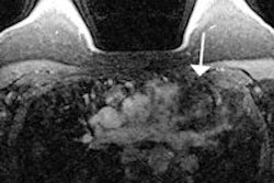
In experienced hands, breast MRI with mammography or ultrasound can help avoid biopsies of suspicious lesions that would otherwise turn out to be false positives, according to a researcher from a leading German institution.
Current clinical practice guidelines from the American College of Radiology (ACR) discourage the use of MRI for working up suspicious findings in mammography and ultrasound. However, MRI improves the positive predictive value of the two technologies, and careful use of the modality could help women avoid unnecessary biopsies.
"Patients don't like to be biopsied, and MRI could help us avoid unnecessary procedures," Dr. Kevin Strobel, from the University of Aachen in Germany, told AuntMinnieEurope.com.
For their study, presented as a poster on 7 September at the American Society of Clinical Oncology 2013 Breast Cancer Symposium in San Francisco, Strobel and colleagues evaluated 2,754 patients who underwent dynamic contrast-enhanced breast MRI between June 2010 and April 2012. Of these, 277 had MRI for further workup of suspicious findings identified with mammography or ultrasound.
All lesions categorized as MRI BI-RADS 4 and 5 underwent biopsy, while lesions categorized as MRI BI-RADS 1 or 2 did not proceed to biopsy, except for women with mammographic calcifications suggesting ductal carcinoma in situ (DCIS). Lesions categorized as MRI BI-RADS 3 had additional short-term follow-up by MRI, ultrasound, and/or mammography. Women who did not undergo biopsy were followed for 12 to 24 months, Strobel said.
For suspicious lesions at mammography, MRI increased positive predictive value as follows:
- Masses: 19.3% for mammography alone to 92.3%
- Asymmetries: 3% for mammography alone to 33.3%
- Architectural distortions: 8.3% for mammography alone to 25%
- Calcifications: 21.1% for mammography alone to 62.5%
For suspicious lesions at ultrasound, MRI increased positive predictive value from 13.5% for ultrasound alone to 71.4% for ultrasound plus MRI for masses, and from 3.3% to 50% for nonmass findings. In four of the 277 patients, MRI showed additional suspicious findings, requiring MR-guided biopsy; one was proved malignant by histology.
Thus far, no patient who avoided biopsy has been diagnosed with invasive cancer or DCIS, according to Strobel.
Even if breast MRI is effective for conclusively working up suspicious findings in mammography and ultrasound, should it be used? Perhaps with a simplified imaging protocol that could reduce time and cost, Strobel said, referring to research presented at the conference by colleague Dr. Christiane Kuhl in which breast MRI acquisition and reading time was cut from 21 minutes to three minutes.
"There's no denying that breast MRI costs more than biopsies," he told AuntMinnieEurope.com. "But we wanted to demonstrate that the modality is viable for this purpose. Perhaps if shorter imaging protocols are developed, using MRI to work up suspicious findings would be less time-consuming and less costly."


![Examples of ultrasound findings and techniques. (A) Images in a 39-year-old male patient with a mass in the left thigh. The mass is heterogeneous on the B-mode US image (compared with the patient in D) and showed increased microvascularity (superb microvascular imaging [SMI]) and shear-wave elastography (SWE) values. Undifferentiated pleomorphic sarcoma was diagnosed at biopsy (with pleomorphic rhabdomyosarcoma in surgical specimen). (B) Images in an 18-year-old male patient with a mass in the left leg. The mass is hypoechoic on the B-mode image, with no other findings suggestive of malignancy. The lesion is in contact with the cortex of the tibia, which is slightly irregular. CT revealed a doubtful anteromedial tibial erosion. The microvascular study demonstrated high vascularization, suggestive of malignancy. Periosteal Ewing sarcoma was diagnosed with both histologic and immunohistochemical confirmation. (C) Images in a 69-year-old female patient with a lump growing on the outside of the left leg. Multiple SWE examinations were performed (please note the high values obtained in the measurements, whereas the color map highlights the stiffness relative to adjacent tissues). SMI showed areas of increased vascularization to target for sampling. Undifferentiated spindle cell sarcoma was diagnosed at biopsy, with residual leiomyosarcoma in the surgical specimen after neoadjuvant therapy. (D) Images in a 56-year-old female patient with a mass in the right thigh. The mass is heterogeneous at both B-mode ultrasound (similar to patient A) and MRI (coronal T2-weighted spectral attenuated inversion recovery [SPAIR]; T1-weighted pre-contrast and postcontrast imaging), which even shows uptake after the administration of paramagnetic contrast material, which is traditionally suggestive of malignancy. Low values at SMI and elastography are suggestive of benignity. Spindle cell lipoma was diagnosed at biopsy, with atypical spindle cell lipomatous tumor in the surgical specimen.](https://img.auntminnieeurope.com/mindful/smg/workspaces/default/uploads/2026/08/images-radiol250278fig2.APCFLSvX6p.jpg?auto=format%2Ccompress&fit=crop&h=100&q=70&w=100)


![Examples of ultrasound findings and techniques. (A) Images in a 39-year-old male patient with a mass in the left thigh. The mass is heterogeneous on the B-mode US image (compared with the patient in D) and showed increased microvascularity (superb microvascular imaging [SMI]) and shear-wave elastography (SWE) values. Undifferentiated pleomorphic sarcoma was diagnosed at biopsy (with pleomorphic rhabdomyosarcoma in surgical specimen). (B) Images in an 18-year-old male patient with a mass in the left leg. The mass is hypoechoic on the B-mode image, with no other findings suggestive of malignancy. The lesion is in contact with the cortex of the tibia, which is slightly irregular. CT revealed a doubtful anteromedial tibial erosion. The microvascular study demonstrated high vascularization, suggestive of malignancy. Periosteal Ewing sarcoma was diagnosed with both histologic and immunohistochemical confirmation. (C) Images in a 69-year-old female patient with a lump growing on the outside of the left leg. Multiple SWE examinations were performed (please note the high values obtained in the measurements, whereas the color map highlights the stiffness relative to adjacent tissues). SMI showed areas of increased vascularization to target for sampling. Undifferentiated spindle cell sarcoma was diagnosed at biopsy, with residual leiomyosarcoma in the surgical specimen after neoadjuvant therapy. (D) Images in a 56-year-old female patient with a mass in the right thigh. The mass is heterogeneous at both B-mode ultrasound (similar to patient A) and MRI (coronal T2-weighted spectral attenuated inversion recovery [SPAIR]; T1-weighted pre-contrast and postcontrast imaging), which even shows uptake after the administration of paramagnetic contrast material, which is traditionally suggestive of malignancy. Low values at SMI and elastography are suggestive of benignity. Spindle cell lipoma was diagnosed at biopsy, with atypical spindle cell lipomatous tumor in the surgical specimen.](https://img.auntminnieeurope.com/mindful/smg/workspaces/default/uploads/2026/08/images-radiol250278fig2.APCFLSvX6p.jpg?auto=format%2Ccompress&fit=crop&h=112&q=70&w=112)