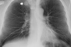
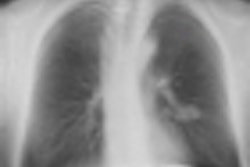
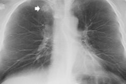
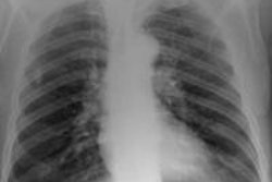
By adding chest digital tomosynthesis in suspected cases of pulmonary lesions, patients can avoid unnecessary CT scans and imaging facilities can lower their expenses, Italian researchers have found.
Radiologists from the University of Trieste sought to evaluate diagnostic imaging costs before and after digital tomosynthesis implementation in patients with suspected thoracic lesions on chest radiography. They presented their results at ECR 2013.
"The detection and characterization of pulmonary lesions represents a challenging task in chest radiography," said lead study author Dr. Emilio Quaia. "This is due to the poor conspicuity of lesions and to the overlapping with anatomical structures. Oblique x-ray views or CT are usually employed to solve doubtful or equivocal cases."
Digital tomosynthesis generates images in different planes throughout a region of interest as the x-ray tube pans around the patient, and this projection data is then reconstructed into 3D images.
Over a period of four years at the university, a total of 465 patients (263 males and 202 females, with a mean age of 72.5 years +/- 11.3 years) with suspected thoracic lesions after chest radiography underwent digital tomosynthesis (Definium 8000, GE Healthcare).
Patient diagnoses
Two observers provided a prospective diagnosis on digital tomosynthesis on a scale of 1 to 5, with 1 as "definitely benign," 3 as "doubtful," and 5 as a "definitely malignant pulmonary lesion." Patients with scores of 1 or 2 received a chest x-ray six months after digital tomosynthesis. Individuals with scores of 3, 4, or 5 underwent CT when digital tomosynthesis identified a definite noncalcified pulmonary lesion, or if the finding was inconclusive.
Among the diagnoses were 60 cases of pulmonary opacities, 26 individuals with pulmonary scars, 47 cases of ground-last opacities and nodules, 32 people with noncalcified solid nodules, 23 subjects with classified solid nodules, 36 patients with pleural plaques, and toured 36 cases of pulmonary pseudolesions.
Of the 465 patients, 338 patients (83%) with scores of 1 or 2 received chest x-ray as their imaging follow-up, while 147 patients (17%) with scores of 3, 4, or 5 were given CT scans.
Quaia and colleagues then calculated unit costs for conventional x-ray, digital tomosynthesis, and CT at their hospital. They compared those expenses after implementing digital tomosynthesis with a second scenario in which only chest x-ray and CT were available to image for pulmonary lesions.
"We also considered only the long-term variable costs, such as personnel, asset depreciation, and maintenance, and the difference between the short-term variable costs," he explained.
Modality comparison
The comparison found that conventional chest x-ray was the least expensive of the imaging choices with unit costs of 15.15 euros ($19.71 U.S.) per scan, compared with unit costs for digital tomosynthesis of 41.55 euros ($54.0 U.S.) per scan. The unit cost for CT without contrast enhancement was 65.35 euros ($84.04) per scan, while contrast-enhanced CT was the most expensive at 113.66 euros ($147.91) per scan. (See chart)
Full unit costs (euros) for imaging modalities
|
Among the 229 patients (49%) who underwent digital tomosynthesis after suspicious findings on chest radiography, digital tomosynthesis discovered 193 thoracic lesions and 36 pleural lesions.
The study also noted in the 12 months prior to implementing digital tomosynthesis, there were 271 patients who presented with suspected thoracic lesions and underwent CT. After digital tomosynthesis became part of the patient workup, the number of patients who received a CT scan decreased to 39 cases. In other words, 232 patents avoided a chest CT exam after digital tomosynthesis results were known.
As for the financial benefit, without digital tomosynthesis, the total annual cost for noncontrast-enhanced CT is approximately 17,891 euros ($23,281 U.S.) and 30,984 euros ($40,319 U.S.) for contrast-enhanced CT using the model developed by Quaia and colleagues.
When digital tomosynthesis was implemented, there were 91 chest x-rays, 130 digital tomosynthesis scans, and 39 CT exams at the hospital. The annual cost for noncontrast-enhanced CT was reduced to 9,329 euros ($12,139 U.S.) and 11,213 euros ($14,591 U.S.) for contrast-enhanced CT. Thus, the annual cost savings for noncontrast-enhanced CT was 8,562 euros ($11,141 U.S.) and 19,771 euros ($25,728 U.S.) for contrast-enhanced CT.
Based on the calculations, the authors recommended that digital tomosynthesis instead of CT may be considered "a first-line imaging technique to rule out pulmonary lesions." In addition, there was a "reduction of CT examination overload and the per-patient diagnostic imaging cost decreased after digital tomosynthesis implementation."