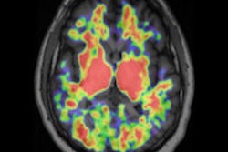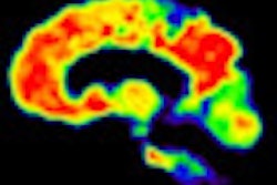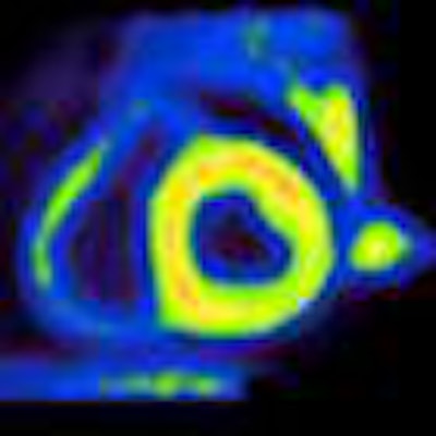
Swedish researchers are expanding the use of PET imaging with carbon-11-labeled Pittsburgh Compound B (PiB) to cardiac amyloidosis, confirming the biomarker's ability to detect amyloid plaque deposits in the heart in a first-of-its-kind study in the February issue of the Journal of Nuclear Medicine.
In patients with cardiac amyloidosis, researchers from Uppsala University found a "significant" increase in PiB as the biomarker bound to amyloid plaque deposits in the heart. PiB-PET also revealed a significant reduction in myocardial blood flow.
"This finding is encouraging in terms of establishing an objective method in a clinical routine to document cardiac amyloidosis in patients for whom cardiac biopsy material is not available," the authors wrote (JNM, February 2013, Vol. 54:2, pp. 213-220). Currently, there is no noninvasive test available for a specific diagnosis of cardiac amyloidosis.
Lead study author is Gunnar Antoni, PhD, from the department of medicinal chemistry at Uppsala University.
PiB for dementia
Until this study, PiB-PET has primarily been known for its utility in detecting biomarkers that are early signs of several forms of dementia. PiB has detected deposits of amyloid plaque in the human brain, which have been associated with the onset of Alzheimer's disease.
Cardiac amyloidosis is a deadly disorder caused by abnormal amyloid deposits in heart tissue. Early diagnosis before structural change to the heart tissue is essential for disease prognosis and also treatment monitoring.
Researchers enrolled 10 patients with a mean age of 66 years, ranging from 48 to 77 years, who were diagnosed with systemic amyloidosis. The study also included five healthy volunteers with a mean age of 64 years, ranging from 54 to 75 years.
 |
| Lef to right: Images of PiB and myocardial blood flow (MBF) in patients with high, intermediate, and partially increased PiB retention and a healthy control. The liver is clearly visible in PiB images of one patient (second from left) and in the healthy control, and is just outside PET field-of-view in two other patients. Liver uptake is due to biliary excretion of PiB and is likely not related to amyloid binding. Images courtesy of the Journal of Nuclear Medicine. |
After a low-dose CT scan, all 15 subjects were given a 32-minute dynamic scan of the heart with an intravenous injection of acetate (10 MBq/kg) using a PET/CT scanner (Discovery ST, GE Healthcare) to measure myocardial blood flow to determine the impact of global and regional perfusion on PiB retention.
At least two hours later, a second low-dose CT scan was performed, followed by a second 25-minute dynamic scan of the heart with an intravenous injection of PiB (6 MBq/kg). Uptake of PiB was measured 15 to 25 minutes after injection.
The study also obtained echocardiography data for all patients for clinical reasons within 206 ± 149 days of the PET scans. For two patients, pre-PET echocardiography was of poor quality or not retrievable, so echocardiography was repeated after PET. Overall, echocardiography findings were characterized as "typical" for patients with cardiac amyloidosis and with normal or mildly decreased left ventricular ejection fraction.
On the other hand, PiB-PET detected signs of pathology. Researchers discovered an "obvious uptake" of PiB in the left ventricle wall of all patients with cardiac amyloidosis, but no uptake of PiB in the healthy volunteers. In half of the patients, PiB was also detected in the right ventricle wall, while nine patients had signs of reversible uptake, with a maximum concentration at 10 to 15 minutes after PiB injection.
Myocardial blood flow was significantly lower in patients with cardiac amyloidosis, but the researchers found no significant correlation between myocardial blood flow and PiB uptake.
Based on the findings, PiB-PET could provide a noninvasive way to show the distribution of amyloid in an organ, according to Antoni and colleagues. The imaging technique may also offer an opportunity to follow and monitor therapy, as amyloid deposits in the heart should decrease with successful therapy.
The researchers plan to advance this research by investigating the potential of quantitative PET-based methods to assess amyloid burden and the dynamics of amyloid turnover in response to direct therapy.
Of the 10 patients with cardiac amyloidosis, five people received melphalan treatment six to 54 months before the PiB-PET scan. The researchers found no significant difference in PiB levels between the groups of patients with and without treatment.
"A possible explanation, that the interindividual variation in PiB retention is larger than the changes in PiB retention caused by melphalan treatment, needs to be further investigated in future studies," they stated.