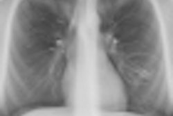
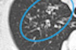
CHICAGO - Ultralow-dose CT is on par with a frontal chest x-ray in terms of dose (0.05 mSv) and gives an accurate diagnosis in pediatric cystic fibrosis patients, Belgian researchers revealed on Monday afternoon at the annual RSNA meeting.
Radiation is always a concern when imaging a patient, but it's even more important for pediatric patients. A chest x-ray is typically used to assess cystic fibrosis, but the modality obviously doesn't offer the same amount of detail as a CT scan. Dr. Caroline Ernst, from University Ziekenhuis in Brussels, and colleagues sought to determine whether ultralow-dose CT could replace chest x-ray altogether for these patients.
In their prospective study, 22 patients underwent ultralow-dose CT. The scans were reconstructed with a model-based iterative reconstruction technique (Veo, GE Healthcare) and randomized and scored using the Bhalla CT scoring system by two radiologists. The ultralow-dose CT scores were compared with Bhalla scores of previous low-dose CT scans obtained in 2009 or 2010.
During her presentation, Ernst clarified that reconstruction is on the model. "It's not like adaptive statistical iterative reconstruction, it's a computer where you put in the information," she said. "We first lowered the dose and you have a lot of image noise, and it's a model that reconstructs images over and over again until an acceptable image [appears]."
The mean CT dose index volume (CTDIvol) for the ultralow-dose CT scans was 0.12 mGy, with a mean estimated effective dose of 0.05 mSv, Ernst said. The mean CTDIvol for the low-dose CT scans was 2.23 mGy, with a mean estimated effective dose of 1.01 mSv.
The mean Bhalla score for the ultralow-dose CT scans was 6.91, compared with 5.65 for the low-dose CT scans. No significant differences were found between the ultralow-dose and low-dose CT scans, Ernst said.
"Our results show that an ultralow-dose CT can be performed with an estimated effective dose of 0.05 mSv, which is in the range of anteroposterior chest dose, and that there is very good agreement in the overall Bhalla score and in the detection and extent of bronchiectasis," she said.
The results show ultralow-dose CT can be performed in pediatric cystic fibrosis patients at a radiation dose that approaches that of a frontal chest radiograph, and that ultralow-dose CT may replace chest x-ray in these patients for a more accurate assessment of bronchiectasis.
"This results in more accurate treatment, but of course more studies have to be done in larger patient groups," Ernst said.


![Examples of ultrasound findings and techniques. (A) Images in a 39-year-old male patient with a mass in the left thigh. The mass is heterogeneous on the B-mode US image (compared with the patient in D) and showed increased microvascularity (superb microvascular imaging [SMI]) and shear-wave elastography (SWE) values. Undifferentiated pleomorphic sarcoma was diagnosed at biopsy (with pleomorphic rhabdomyosarcoma in surgical specimen). (B) Images in an 18-year-old male patient with a mass in the left leg. The mass is hypoechoic on the B-mode image, with no other findings suggestive of malignancy. The lesion is in contact with the cortex of the tibia, which is slightly irregular. CT revealed a doubtful anteromedial tibial erosion. The microvascular study demonstrated high vascularization, suggestive of malignancy. Periosteal Ewing sarcoma was diagnosed with both histologic and immunohistochemical confirmation. (C) Images in a 69-year-old female patient with a lump growing on the outside of the left leg. Multiple SWE examinations were performed (please note the high values obtained in the measurements, whereas the color map highlights the stiffness relative to adjacent tissues). SMI showed areas of increased vascularization to target for sampling. Undifferentiated spindle cell sarcoma was diagnosed at biopsy, with residual leiomyosarcoma in the surgical specimen after neoadjuvant therapy. (D) Images in a 56-year-old female patient with a mass in the right thigh. The mass is heterogeneous at both B-mode ultrasound (similar to patient A) and MRI (coronal T2-weighted spectral attenuated inversion recovery [SPAIR]; T1-weighted pre-contrast and postcontrast imaging), which even shows uptake after the administration of paramagnetic contrast material, which is traditionally suggestive of malignancy. Low values at SMI and elastography are suggestive of benignity. Spindle cell lipoma was diagnosed at biopsy, with atypical spindle cell lipomatous tumor in the surgical specimen.](https://img.auntminnieeurope.com/mindful/smg/workspaces/default/uploads/2026/08/images-radiol250278fig2.APCFLSvX6p.jpg?auto=format%2Ccompress&fit=crop&h=100&q=70&w=100)


![Examples of ultrasound findings and techniques. (A) Images in a 39-year-old male patient with a mass in the left thigh. The mass is heterogeneous on the B-mode US image (compared with the patient in D) and showed increased microvascularity (superb microvascular imaging [SMI]) and shear-wave elastography (SWE) values. Undifferentiated pleomorphic sarcoma was diagnosed at biopsy (with pleomorphic rhabdomyosarcoma in surgical specimen). (B) Images in an 18-year-old male patient with a mass in the left leg. The mass is hypoechoic on the B-mode image, with no other findings suggestive of malignancy. The lesion is in contact with the cortex of the tibia, which is slightly irregular. CT revealed a doubtful anteromedial tibial erosion. The microvascular study demonstrated high vascularization, suggestive of malignancy. Periosteal Ewing sarcoma was diagnosed with both histologic and immunohistochemical confirmation. (C) Images in a 69-year-old female patient with a lump growing on the outside of the left leg. Multiple SWE examinations were performed (please note the high values obtained in the measurements, whereas the color map highlights the stiffness relative to adjacent tissues). SMI showed areas of increased vascularization to target for sampling. Undifferentiated spindle cell sarcoma was diagnosed at biopsy, with residual leiomyosarcoma in the surgical specimen after neoadjuvant therapy. (D) Images in a 56-year-old female patient with a mass in the right thigh. The mass is heterogeneous at both B-mode ultrasound (similar to patient A) and MRI (coronal T2-weighted spectral attenuated inversion recovery [SPAIR]; T1-weighted pre-contrast and postcontrast imaging), which even shows uptake after the administration of paramagnetic contrast material, which is traditionally suggestive of malignancy. Low values at SMI and elastography are suggestive of benignity. Spindle cell lipoma was diagnosed at biopsy, with atypical spindle cell lipomatous tumor in the surgical specimen.](https://img.auntminnieeurope.com/mindful/smg/workspaces/default/uploads/2026/08/images-radiol250278fig2.APCFLSvX6p.jpg?auto=format%2Ccompress&fit=crop&h=112&q=70&w=112)