Adding digital breast tomosynthesis (DBT) to digital mammography boosts accuracy in finding soft-tissue abnormalities, researchers report in the October issue of Clinical Radiology. And that can only help reduce both healthcare costs and patient anxiety.
"The accuracy of conventional mammography is limited by anatomical noise resulting from the superimposition of normal structures," wrote researchers from King's College Hospital in London. "In clinical practice, this may affect both sensitivity and specificity. ... [DBT] has the potential to improve the accuracy of mammography by enabling the reader to view x-ray images of the breast tissue as a series of thin reconstructed sections, so overcoming the problem of overlying tissues on conventional two-dimensional images."
Lead author Dr. Michael Michell and colleagues included 738 women in the study. The women had been recalled for assessment following routine screening with film-screen mammography; between January 2009 and July 2010, each underwent bilateral, two-view full-field digital mammography (FFDM) and two-view DBT.
Readers scored each lesion separately for probability of malignancy on film-screen mammography, FFDM, and then DBT. The scores were compared with the presence or absence of malignancy based on the final histopathology outcome (Clinical Radiology, October 2012, Vol. 67:10, pp. 976-981).
In all, 204 out of 759 lesions found were malignant; 147 (72%) were invasive and 57 (28%) were in situ.
Michell's team performed receiver operator characteristics (ROC) analysis to assess and compare the diagnostic accuracy of the three imaging methods, calculating the area under the curve (AUC):
- Film-screen mammography alone had an AUC of 0.7882.
- FFDM and film-screen mammography combined produced an AUC of 0.8949.
- Adding DBT boosted FFDM and film-screen mammography's AUC to 0.9671.
The difference in AUC between DBT and FFDM with film-screen mammography combined was 0.0722 (p = 0.0001), and the difference in AUC between DBT and film-screen mammography alone was 0.1789 (p = 0.0001).
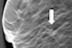
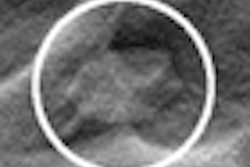
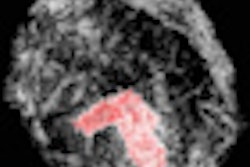
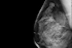
FFDM vs. DBT for histology-proven malignant lesions
|
DBT allowed the readers to classify ambiguous lesions found on FFDM more accurately, the authors wrote. The technology also found six additional cancers that film-screen mammography and FFDM missed.
And increased accuracy improves healthcare, Michell and colleagues noted.
"Better visualization of normal and benign features may improve the ability of the film reader to confidently diagnose cases without malignancy and thereby reduce the recall rate, with savings in resources for the healthcare system and fewer women suffering the anxiety and inconvenience associated with recall," they wrote.


![Examples of ultrasound findings and techniques. (A) Images in a 39-year-old male patient with a mass in the left thigh. The mass is heterogeneous on the B-mode US image (compared with the patient in D) and showed increased microvascularity (superb microvascular imaging [SMI]) and shear-wave elastography (SWE) values. Undifferentiated pleomorphic sarcoma was diagnosed at biopsy (with pleomorphic rhabdomyosarcoma in surgical specimen). (B) Images in an 18-year-old male patient with a mass in the left leg. The mass is hypoechoic on the B-mode image, with no other findings suggestive of malignancy. The lesion is in contact with the cortex of the tibia, which is slightly irregular. CT revealed a doubtful anteromedial tibial erosion. The microvascular study demonstrated high vascularization, suggestive of malignancy. Periosteal Ewing sarcoma was diagnosed with both histologic and immunohistochemical confirmation. (C) Images in a 69-year-old female patient with a lump growing on the outside of the left leg. Multiple SWE examinations were performed (please note the high values obtained in the measurements, whereas the color map highlights the stiffness relative to adjacent tissues). SMI showed areas of increased vascularization to target for sampling. Undifferentiated spindle cell sarcoma was diagnosed at biopsy, with residual leiomyosarcoma in the surgical specimen after neoadjuvant therapy. (D) Images in a 56-year-old female patient with a mass in the right thigh. The mass is heterogeneous at both B-mode ultrasound (similar to patient A) and MRI (coronal T2-weighted spectral attenuated inversion recovery [SPAIR]; T1-weighted pre-contrast and postcontrast imaging), which even shows uptake after the administration of paramagnetic contrast material, which is traditionally suggestive of malignancy. Low values at SMI and elastography are suggestive of benignity. Spindle cell lipoma was diagnosed at biopsy, with atypical spindle cell lipomatous tumor in the surgical specimen.](https://img.auntminnieeurope.com/mindful/smg/workspaces/default/uploads/2026/08/images-radiol250278fig2.APCFLSvX6p.jpg?auto=format%2Ccompress&fit=crop&h=100&q=70&w=100)


![Examples of ultrasound findings and techniques. (A) Images in a 39-year-old male patient with a mass in the left thigh. The mass is heterogeneous on the B-mode US image (compared with the patient in D) and showed increased microvascularity (superb microvascular imaging [SMI]) and shear-wave elastography (SWE) values. Undifferentiated pleomorphic sarcoma was diagnosed at biopsy (with pleomorphic rhabdomyosarcoma in surgical specimen). (B) Images in an 18-year-old male patient with a mass in the left leg. The mass is hypoechoic on the B-mode image, with no other findings suggestive of malignancy. The lesion is in contact with the cortex of the tibia, which is slightly irregular. CT revealed a doubtful anteromedial tibial erosion. The microvascular study demonstrated high vascularization, suggestive of malignancy. Periosteal Ewing sarcoma was diagnosed with both histologic and immunohistochemical confirmation. (C) Images in a 69-year-old female patient with a lump growing on the outside of the left leg. Multiple SWE examinations were performed (please note the high values obtained in the measurements, whereas the color map highlights the stiffness relative to adjacent tissues). SMI showed areas of increased vascularization to target for sampling. Undifferentiated spindle cell sarcoma was diagnosed at biopsy, with residual leiomyosarcoma in the surgical specimen after neoadjuvant therapy. (D) Images in a 56-year-old female patient with a mass in the right thigh. The mass is heterogeneous at both B-mode ultrasound (similar to patient A) and MRI (coronal T2-weighted spectral attenuated inversion recovery [SPAIR]; T1-weighted pre-contrast and postcontrast imaging), which even shows uptake after the administration of paramagnetic contrast material, which is traditionally suggestive of malignancy. Low values at SMI and elastography are suggestive of benignity. Spindle cell lipoma was diagnosed at biopsy, with atypical spindle cell lipomatous tumor in the surgical specimen.](https://img.auntminnieeurope.com/mindful/smg/workspaces/default/uploads/2026/08/images-radiol250278fig2.APCFLSvX6p.jpg?auto=format%2Ccompress&fit=crop&h=112&q=70&w=112)