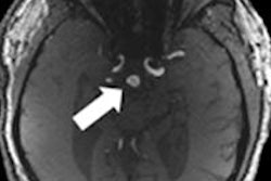
Standard head movements made while exposed to electromagnetic fields produced by a 7-tesla MRI scanner seem to temporarily lower concentration and visuospatial awareness, according to a study published online on Monday in Occupational and Environmental Medicine.
Researchers from the Institute for Risk Assessment Sciences at Utrecht University in the Netherlands wrote that the effects from the scanner were particularly noticeable when participants were asked to use high levels of working memory. The findings may have implications for surgeons and other healthcare providers working near a high-field MRI scanner.
In the study, 31 volunteers made standard head movements within the static magnetic field of a 7-tesla MRI unit at exposure levels of zero, 0.5 (medium), and 1 (high) tesla in a random order one week apart.
After each exposure level, the volunteers were given 12 timed cognitive tasks to test the kinds of skills a surgeon or clinician might need to perform within the vicinity of an MRI scanner. The tasks included visual tracking and movement and general functions such as attention, concentration, and working memory.
A total of 30 volunteers completed all three sessions. The results showed that the more general functions such as attention and concentration, as well as visuospatial awareness, were significantly affected at 0.5 and 1 tesla, compared with actions performed at 0 tesla.
For complex mental tasks, reaction and disengagement times were longer, varying from 5% to 21% when exposed to greater levels of magnet strength.
While nonverbal memory did not seem to be affected by the degree of tesla, the study showed a decline in verbal memory, although the result had only borderline significance. At the highest level of exposure, volunteers also experienced some physical symptoms including metallic taste in the mouth among 12 people, dizziness in six, and headaches in five. One person reported nausea.
"The exact implications and mechanisms of these subtle acute effects in [practice] remain unclear," wrote Dr. Hans Kromhout and colleagues. "To date, mainly health and safety concerns for patients have been evaluated, but possible consequences are particularly important for professionals ... cleaners, and MRI engineers, since they are repeatedly exposed to static magnetic fields."


![Examples of ultrasound findings and techniques. (A) Images in a 39-year-old male patient with a mass in the left thigh. The mass is heterogeneous on the B-mode US image (compared with the patient in D) and showed increased microvascularity (superb microvascular imaging [SMI]) and shear-wave elastography (SWE) values. Undifferentiated pleomorphic sarcoma was diagnosed at biopsy (with pleomorphic rhabdomyosarcoma in surgical specimen). (B) Images in an 18-year-old male patient with a mass in the left leg. The mass is hypoechoic on the B-mode image, with no other findings suggestive of malignancy. The lesion is in contact with the cortex of the tibia, which is slightly irregular. CT revealed a doubtful anteromedial tibial erosion. The microvascular study demonstrated high vascularization, suggestive of malignancy. Periosteal Ewing sarcoma was diagnosed with both histologic and immunohistochemical confirmation. (C) Images in a 69-year-old female patient with a lump growing on the outside of the left leg. Multiple SWE examinations were performed (please note the high values obtained in the measurements, whereas the color map highlights the stiffness relative to adjacent tissues). SMI showed areas of increased vascularization to target for sampling. Undifferentiated spindle cell sarcoma was diagnosed at biopsy, with residual leiomyosarcoma in the surgical specimen after neoadjuvant therapy. (D) Images in a 56-year-old female patient with a mass in the right thigh. The mass is heterogeneous at both B-mode ultrasound (similar to patient A) and MRI (coronal T2-weighted spectral attenuated inversion recovery [SPAIR]; T1-weighted pre-contrast and postcontrast imaging), which even shows uptake after the administration of paramagnetic contrast material, which is traditionally suggestive of malignancy. Low values at SMI and elastography are suggestive of benignity. Spindle cell lipoma was diagnosed at biopsy, with atypical spindle cell lipomatous tumor in the surgical specimen.](https://img.auntminnieeurope.com/mindful/smg/workspaces/default/uploads/2026/08/images-radiol250278fig2.APCFLSvX6p.jpg?auto=format%2Ccompress&fit=crop&h=100&q=70&w=100)


![Examples of ultrasound findings and techniques. (A) Images in a 39-year-old male patient with a mass in the left thigh. The mass is heterogeneous on the B-mode US image (compared with the patient in D) and showed increased microvascularity (superb microvascular imaging [SMI]) and shear-wave elastography (SWE) values. Undifferentiated pleomorphic sarcoma was diagnosed at biopsy (with pleomorphic rhabdomyosarcoma in surgical specimen). (B) Images in an 18-year-old male patient with a mass in the left leg. The mass is hypoechoic on the B-mode image, with no other findings suggestive of malignancy. The lesion is in contact with the cortex of the tibia, which is slightly irregular. CT revealed a doubtful anteromedial tibial erosion. The microvascular study demonstrated high vascularization, suggestive of malignancy. Periosteal Ewing sarcoma was diagnosed with both histologic and immunohistochemical confirmation. (C) Images in a 69-year-old female patient with a lump growing on the outside of the left leg. Multiple SWE examinations were performed (please note the high values obtained in the measurements, whereas the color map highlights the stiffness relative to adjacent tissues). SMI showed areas of increased vascularization to target for sampling. Undifferentiated spindle cell sarcoma was diagnosed at biopsy, with residual leiomyosarcoma in the surgical specimen after neoadjuvant therapy. (D) Images in a 56-year-old female patient with a mass in the right thigh. The mass is heterogeneous at both B-mode ultrasound (similar to patient A) and MRI (coronal T2-weighted spectral attenuated inversion recovery [SPAIR]; T1-weighted pre-contrast and postcontrast imaging), which even shows uptake after the administration of paramagnetic contrast material, which is traditionally suggestive of malignancy. Low values at SMI and elastography are suggestive of benignity. Spindle cell lipoma was diagnosed at biopsy, with atypical spindle cell lipomatous tumor in the surgical specimen.](https://img.auntminnieeurope.com/mindful/smg/workspaces/default/uploads/2026/08/images-radiol250278fig2.APCFLSvX6p.jpg?auto=format%2Ccompress&fit=crop&h=112&q=70&w=112)