A new survey suggests that a widespread lack of awareness of radiation doses and the potential risks of ionizing radiation exists among junior doctors, and has confirmed the need to educate current and future referring doctors.
A total of 60 foundation doctors in North-West London completed a questionnaire designed to test their knowledge of radiation doses associated with common diagnostic imaging procedures. They were also asked to specify the certainty of their answers. Other questions focused on identifying which investigations emit ionizing radiation. The trainees received no prior tutorials or lectures on this subject.
A foundation doctor is a medical practitioner in the U.K. undertaking the Foundation Programme, which is a two-year, general postgraduate medical training program that forms the bridge between medical school and specialist/general practice training.
"The increasing use of diagnostic imaging studies has given rise to growing fears over the risks associated with the high levels of radiation exposures," noted Dr. Sheena Patel, a surgical trainee at St. Mark's Hospital, London, who presented the findings from the survey at the 2012 UK Radiological Congress (UKRC). "Many studies have also raised concerns over the limited awareness of these risks amongst medical students and referring doctors."
The trainees were given a list of investigations and asked which involved the use of ionizing radiation. For both abdominal x-rays (AXRs) and intravenous urograms (IVUs), the number of incorrect answers was nine (15%). For barium studies, 10 trainees (17%) gave the wrong answer, whereas 14 respondents (23%) gave the incorrect answer for mammography, 15 (25%) were wrong about angiograms, 18 (30%) were wrong about ventilation/perfusion (V/Q) scans, 27 (45%) were wrong about PET scans, six (10%) were wrong about MRI, and seven (12%) were wrong about CT.
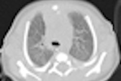
For radiation doses from different investigations, the answers were classified as "underestimate," "correct," or "overestimate." A total of 13 trainees overestimated the radiation dose of AXRs, and the remaining 47 were correct. For IVUs, 28 underestimated the dose, eight overestimated the dose, and 24 were correct. For head CT scans, seven underestimated the dose, 43 overestimated the dose, and 10 were correct. For CT scans of the abdomen and pelvis, 27 underestimated the dose, and 33 were correct.
Overall, of the correctly answered questions, 54% were guesses, 10% were certain, and 36% were somewhat certain. A quarter of trainees correctly identified the lifetime risk (1 in 2000) of inducing a fatal cancer from an abdominal CT, and 58% underestimated this risk.
"Since foundation doctors are responsible for organizing and requesting various diagnostic imaging studies, it is crucial they are aware of radiation exposures associated with these investigations," Patel stated.