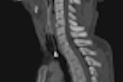
A basic knowledge of ballistics is essential to enable detection and interpretation of the lesions resulting from gunshot wounds in the abdomen and thorax, and contrast-enhanced CT is the best examination to perform in stable patients, French researchers have found.
"CT must image one body region above and one body region below any wound, as well as the region involved when searching for foreign bodies. CT must be performed without and with contrast media injection to search active bleeding and organ lesions," noted Dr. Victoire Cartier and colleagues from the department of radiology at Centre Hospitalier Universitaire d'Angers, France, in an e-poster at the 2012 European Society of Gastrointestinal and Abdominal Radiology (ESGAR) congress in Edinburgh, U.K.
Multiplane reconstructed images and 3D reconstructions are helpful to determine wound path trajectories and also entrance and exit wounds, as well as to understand the lesions, they added.
Abdominal and thoracic gunshot injuries are often secondary to suicide and hunting accidents, and require an instant prognosis. CT offers a fast, noninvasive analytical tool for hemodynamically stable patients, while the other patients should be operated on immediately, Cartier stated. The modality allows identification of organ lesions to plan surgical procedures or conservative treatment.
"An understanding of general ballistic principles is of major importance to guide clinical management of patients with gunshot injuries," the authors wrote. "Lesions depend on projectile type and resilience of injured tissues. Knowledge of the gun, and especially the projectile, permits us to better understand and visualize the lesions."
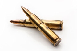
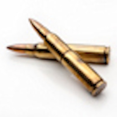
There are two different types of firearms -- low- and high-velocity -- and the projectile will depend on the firearm used. The most common projectiles are bullets and pellets, and for the ballistic data, three elements have to be considered: the entrance wound, the exit wound (which is often diametrically opposite to the entrance wound), and the path of the projectile. There may be no exit wound when the projectile is still in the body, they explained.
When a bullet enters the body, energy is transmitted to tissues, creating two areas of projectile-tissue interaction: the permanent and the temporary cavity. The degree of tissue disruption caused by a projectile is related to the size of the temporary versus permanent cavity. The extent of cavitation is related to the characteristics of the projectile, according to Cartier and colleagues. Furthermore, tissue density and elasticity influence the wounding capacity of a bullet. Dense tissues -- liver, spleen, kidney, full bladder, and stomach -- distort the path of a bullet and cause increased tissue destruction.
"Bony involvement may cause additional injuries, producing bony fragments, which act as minor projectiles. When projectiles are pellets, lesions depend on the range, which is the distance between the muzzle of the weapon and the victim," they noted. "Thoracic injuries can be associated with heart and larger blood vessels damages, hematothorax, pneumothorax, contusions, mediastinal lesions, and rib fractures. Abdominal injuries are commonly associated with pneumoperitoneum, bleeding, and organ lesions (stomach, bowel, kidney, liver, and spleen)."