Effective treatment of acute small-bowel disorders depends critically on early and accurate diagnosis, as well as radiologists' familiarity with the CT appearances of different pathologies, a leading group of researchers from Portugal have stressed.
"CT is increasingly being used as a tool for evaluation of patients with acute symptoms of intestinal disease because of its wide availability and assessment of other extraintestinal abnormalities," noted Dr. Lara Batista, from the department of radiology at Centro Hospitalar Vila Nova de Gaia, during the 2012 European Society of Gastrointestinal and Abdominal Radiology (ESGAR) congress in Edinburgh, U.K. "A variable pattern of intestinal wall morphologic and enhancement abnormalities can be identified with CT in small intestine diseases, including inflammatory, vascular, mechanic, traumatic and tumoral disorders."
Acute small-bowel disorders represent a rare but important condition seen in the emergency department and require an urgent therapeutic decision, she stated. The differential diagnosis includes a wide variation of diseases. Abdominal radiographs and ultrasound are often insufficient, whereas CT allows a quick and accurate evaluation of patients, and not only studies the small intestine, but also the mesentery, adjacent solid organs and the peritoneum, and retroperitoneum.
Acute gastroenteritis is one of the most common causes of small-bowel disorders, and though imaging is often not needed, it can show mural thickening, small-bowel luminal narrowing, and in some cases ulcerations and perforation, according to Batista and her colleagues. In gastrointestinal infection by mycobacterium tuberculosis, the ileocecal area is the most affected, as in Crohn's disease, and it can cause small-bowel obstruction.
"CT may demonstrate thickening of folds, inflammatory heterogeneous mass, and large nodes with hypoattenuating centers due to necrosis. Sometimes it is necessary to obtain an image to exclude the presence of an abscess," she said. "Inflammatory bowel disease is one of the most common causes of abdominal abscess. Patients usually present with fever and leukocytosis. CT is diagnostic and shows a loculated fluid collection outside the bowel lumen, with peripheral rim enhancement and sometimes gas."
In Crohn's disease, the CT findings that suggest active disease include bowel wall thickening (usually more than 1 cm) with mural stratification and mucosa and mural hyperenhancement (target-like, "double halo" appearance), involvement of the mesentery (stranding of the mesenteric fat, "creeping fat," phlegmon, and abscess formation), fistula (ileocolonic, ileocecal, enterocutaneous, enterovesical, colovesical) and sinus tract formation, increased separation of ileal vasa recta, vascular jejunization of the ileum ("comb-sign"), and lymphadenopathy.
In the long-standing stenotic phase, some patients have recurrent episodes of partial obstruction of the bowel due to strictures, areas of ulceration and spasm and secondary to adhesions, incisional hernias and postoperative strictures, according to Batista. In the active disease, luminal narrowing due to acute inflammatory process can also lead to obstruction, and CT can determine the site, level, and the cause of the obstruction. These patients have an increased prevalence of tumors, such as carcinomas of the small and large bowel. Recurrences after resection of disease bowel commonly occur at the anastomic portion.
Small-bowel diverticula are rare, but are more often seen in the duodenum. On CT studies, they appear as a blind-ending tubular structure of variable size, with air and fluid, located in the abdomen or pelvis and attached to the antimesenteric border of the ileum. Wall thickening and mesenteric soft-tissue stranding suggest acute inflammation.
In cases of radiation enteritis, CT provides useful information about small-bowel damage. CT findings include long segments of bowel with wall thickening and mucosal hyperenhancement, stranding of themesenteric fat, hyperemia of mesenteric vessels, and separation of bowel loops due to mesenteric edema. The affected folds can appear fixed and angulated, and fibrosis can occur, leading to tapered strictures within long segments of the bowel. Fistulization can also occur between pelvic organs, she noted.
"Sometimes small-bowel obstruction develops because of adhesions and fibrotic changes in the mesentery and, in rare cases, because of luminal narrowing and dismotility caused by radiation serositis. Ileal loops are the most affected," Batista pointed out. "CT demonstrates mural thickening with consequent luminal narrowing, abnormal enhancement of the bowel wall, mesenteric retraction, and angular bowel wall caused by adhesions. Inflammation, intussusception, bleeding, ulceration, enterolith formation, volvulus, and perforation are some of the possible complications."
CT is the procedure of choice when bowel ischemia is suspected and may help to determine the cause, such as vascular occlusion due to thrombus, embolus, or arteriosclerotic disease, compression secondary to bowel occlusion, vascular tumor invasion, plus the possible complications. It is essential to obtain a biphasic acquisition, the arterial phase beginning at 25-30 seconds after contrast injection and the portal venous phase at 60 seconds, she stated.
Traumatic injuries to the small bowel are rare and challenging. Jejunum and ileum are the most usually affected bowel parts by blunt abdominal trauma, often near the point of fixation (ileocecal valve, ligament of Treitz). The second and third parts are the most affected segments of the duodenum. CT is the modality of choice in hemodynamically stable patients for evaluating bowel and mesentery injuries because of its high accuracy.
Carcinoid tumor is the most common primary neoplasm of the small bowel, and it is commonly a solitary lesion, but can be multiple in 30% of cases, Batista explained. CT can demonstrate the tethering of small-bowel folds, encasement and luminal narrowing, retroperitoneal adenopathy and hypervascular liver metastases that can sometimes have central necrosis. Regional metastases or lymph nodes are commonly seen in the mesentery and have a stellate appearance with a central mass and calcifications that separate the small-bowel loops.
Adenocarcinoma is most commonly found in the proximal small bowel, particularly in the duodenum, and it has a poor prognosis because of high incidence of lymphatic and hematogenous spread by the time of diagnosis, she added. Symptoms are nonspecific and include abdominal pain, bleeding, anemia, and obstruction. When a small-bowel adenocarcinoma presents as obstruction, often it is in an advanced state and CT shows an asymmetric thickening of the bowel walls at the transition point.
Small-bowel obstruction is a frequent clinical situation in the emergency department, and CT can reveal the site, level, cause, and severity of obstruction and to display the presence of strangulation and signs of threatened bowel viability. It may not require oral contrast because retained intraluminal fluid provides sufficient negative contrast, allowing evaluation of the extramural areas. Intravenous contrast administration is important, especially when ischemia is suspected, noted Batista and her colleagues.
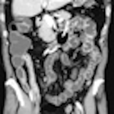
Editor's note: The CT image on our home page shows a small-bowel enteroclysis of a patient with Crohn's disease extended throughout the ileum. Image courtesy of Dr. Gian Rollandi, Ospedali Galliera, Genoa, Italy.