Diffusion-weighted MRI (DWI) and apparent diffusion coefficient (ADC) measurements help increase the specificity of breast MRI to more than 90% and provide faster imaging times in a noninvasive, nonionizing radiation way, concludes a new meta-analysis from France.
Researchers also found that lesion detection is better at 3-tesla MRI than at 1.5-tesla MRI, and ADC measurements are accurate for both masses and areas of nonmass-like enhancement, regardless of lesion size.
Lead study author is Dr. Isabelle Thomassin-Naggara from the department of radiology at Hôpital Tenon, Assistance Publique Hôpitaux de Paris, and Université Pierre et Marie Curie in Paris. Her article-in-press was published online on 31 May in the European Journal of Radiology.
As previous studies have noted, routine lesion detection via breast MRI is aided by dynamic enhancement with contrast agents, which traditionally yield diagnostic sensitivity of 94% to 99%. The trade-off, however, is lower specificity, which can range from 37% to 86%.

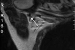
 Invasive ductal carcinoma with positive axillary lymphadenopathy. The detection of malignant masses is relatively easy using DWI for both index tumor and axillary positive lymph node. ADC values of the index tumor and positive homolateral lymph node were less than 0.9. This case illustrates the usefulness of DWI to detect both index tumor and positive lymphadenopathy. Images courtesy of Dr. Isabelle Thomassin-Naggara.
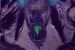
With the development of advanced MRI techniques, such as diffusion-weighted imaging, the potential exists for improving lesion detection and characterization in the breast, Thomassin-Naggara noted.
DWI, for example, calculates the movement of water molecules, while lesion characterization is based on the detection of changes in water molecule ability. "Therefore, in addition to contrast enhancement-based characterization, measurement of the motion of water molecules in DWI provides an additional feature for lesion characterization that may further increase the specificity of MRI for classifying breast lesions," the authors wrote.
3 tesla vs. 1.5 tesla
Thomassin-Naggara and colleagues found only one study that compared 1.5- and 3-tesla MRI magnet strengths. Matsuoka and colleagues evaluated 16 lesions and 13 patients who underwent MRI exams with both magnet strengths. The study found no significant difference for ADC values between the two MRI devices, the lesions less than 10 mm in size were more clearly visible and better delineated at 3 tesla. However, researchers noted that the exam protocol varied in this study, with thinner image slices acquired with 3 tesla compared with 1.5 tesla (Radiation Medicine, January 2008, Vol. 28:1, pp.15-20).
Further studies are necessary to confirm these preliminary results, Thomassin-Naggara and colleagues wrote.
ADC measurements
The authors also cited previous research, which found no difference in ADC measurements acquired by single-shot spin-echo echo-planar imaging (EPI-DWI) or half-Fourier acquisition single-shot turbo spin-echo (HASTE-MRI) protocols. Both techniques resulted in similar diagnostic value in determining benign and malignant lesions, according to Baltzer and colleagues, but EPI-DWI outperformed HASTE-DWI in lesion visibility, 89% to 78%, respectively (Eur Radiol, July 2009, Vol. 19:7, pp. 1612-1620).
A study by Takahara et al indicated that breast lesion detection could be further optimized through DWI whole-body imaging, which was developed to perform whole-body MRI for malignancies (Radiation Medicine, 2004, Vol. 22:4, pp. 275-282).
Previously, Stadlbauer et al found that DWI whole-body imaging was superior to DWI in the visualization of malignant and benign lesions with better detection of 96% compared with 72%, respectively. In addition, ADC values were generally higher for DWI whole-body imaging than for DWI due to the inversion pulse used in this MRI technique. The conclusion was that overall diagnostic performance of ADC values to distinguish benign from malignant lesions was not significantly different between the two techniques (European Radiology, 2009, Vol. 19: 10, pp. 2349-2356).
Benign vs. malignant
In 2009, Kim and colleagues found the cancer detection rate for DWI was 92% in their study of 67 tumors, ranging in size from 0.3 to 1.1 cm. DWI was able to detect mammographically and clinically occult breast carcinomas, including lesions seen on MRI as foci or areas of nonmass-like enhancement (Journal of Magnetic Resonance Imaging, September 2009, Vol. 30:3, pp. 615-620).
The performance of DWI to discriminate between benign and latent tumors also is very good, according to a 2009 study by Yili and colleagues. The research had accuracy as high as 97% in characterizing benign and malignant lesions (BMC Cancer, January 14, 2009, Vol. 9:18).
In evaluating ADC's ability to predict malignancy, Tsushima and colleagues calculated sensitivity of 89% and specificity of 77% (J Magn Reson Imaging, August 2009, Vol. 30:2, pp. 249-255). The results were comparable to a meta-analysis done by Peters et al, which determined contrast-enhancement breast MRI achieved sensitivity of 90% and specificity of 72% (Radiology, January 2008, Vol. 246:1, pp. 116-124).
"While the overall diagnostic performance of contrast-enhanced breast MRI and DWI with ADC measurements may be similar, the latter has the advantage of not using contrast material," Thomassin-Naggara noted.
Overall ADC measurements offer quantitative assessments of breast lesions' characteristics, which result in increased specificity of breast MRI to more than 90%, the French researchers concluded. "Diffusion-weighted imaging is rapid (mean time = 200 seconds), noninvasive, does not involve ionizing radiation, nor does it require injection of a contrast agent," they added.