A novel beam's eye view x-ray imaging system could be used to monitor tumor motion during scanned proton therapy, researchers from a Swiss proton therapy center told attendees at the annual meeting of the European Society for Radiotherapy and Oncology (ESTRO) held in Barcelona, Spain.
The Paul Scherrer Institut (PSI) in Villigen, Switzerland, was a pioneer in the development of pencil-beam scanned proton therapy. The site's newest proton gantry -- Gantry 2 -- is dedicated to fast scanning, offering the ability to perform ultrafast volumetric target repainting and intensity-modulated proton therapy. But while active scanned proton delivery offers many advantages over passively scattered protons, the technique is also particularly sensitive to geometric and temporal variations.
To address this issue, PSI has equipped Gantry 2 with a novel x-ray imaging system that acquires beam's eye view images in the same direction as the treatment field. The aim is to use this beam's eye view imaging system to monitor tumor motion during scanned beam delivery, and ultimately to enable real-time treatment adjustments.
Ye Zhang, a doctoral student at PSI, described the latest developments in this project.
The beam's eye view imaging system, she explained, uses an x-ray tube mounted on the back of the final 90° bending magnet, with the x-rays passing through a hole in the return yoke. The EPID detector panel is mounted under the patient bed on an extractable support. The source-to-detector distance is 444 cm and the source-to-isocenter distance is 395 cm, providing a field size at the isocenter of 20 x 25 cm.
"So how can we use this innovative imaging system to guide scanned proton therapy?" Zhang asked. Tumor motion during treatment, such as that due to respiration, for example, is often addressed by expanding the clinical target volume to an internal target volume that encompasses the motion envelope. The ultimate aim of image-guided treatment, explained Zhang, is to reduce the size of this internal target volume.
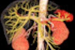
The PSI team has developed algorithms that perform automatic recognition of multiple fiducial markers and segmentation of diaphragm images. Zhang noted that, in simulation studies, both diaphragm and marker motion could be observed and extracted, implying that this information could be used to guide gating or breath-hold during treatment.
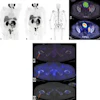
To examine the system's potential for intrafractional tumor motion tracking, the team generated time-resolved digitally reconstructed radiographs from simulated 4D CT datasets (static CT images with motion information from 4D MRI data) of liver cancer patients. These contained motion information over a relatively long time, including breath-hold and irregular breathing patterns. The digitally reconstructed radiographs, an approximation of the expected beam's eye view images, were used to simulate a patient scenario, using the actual system geometry taken from calibration results.
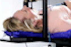
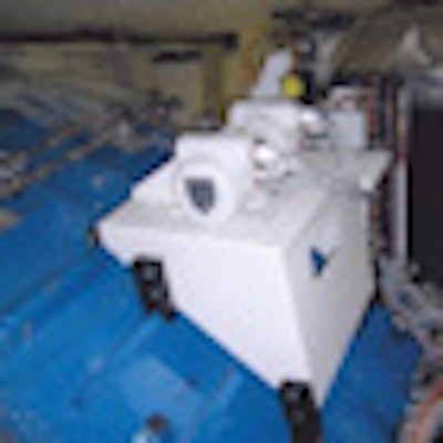
 The beam's eye view configuration. The upper photo shows the x-ray tube; the lower photo shows the EPID imager and the phantom used for calibration of the imaging system. Image courtesy of Paul Scherrer Institut.
In addition, from commissioning results of the beam's eye view imaging system, the accuracy of positional information obtained from the beam's eye view image was within 0.5 mm for all possible gantry angles.
In the future, said Zhang, the system could potentially be used for "slow tracking," or motion management of the target's baseline drift, by performing beam's eye view-guided breath-hold combined with rescanning. This process involves recording an initial surface image to gauge the respiratory signal, performing breath hold at the correct phases and acquiring the beam's eye view image. This image is compared to digitally reconstructed radiographic images calculated from the treatment plan and the positional variation is determined. If this variation is within the specified tolerance, the beam is switched on; if not, the above process is repeated.
"Our beam's eye view imaging system is functional and our online motion extraction algorithms are working for both fiducial marker recognition and diaphragm segmentation," Zhang concluded. "This concept of beam's eye view image-guided 'slow tracking' strategy is feasible, but still needs more work."
© IOP Publishing Limited. Republished with permission from medicalphysicsweb, a community website covering fundamental research and emerging technologies in medical imaging and radiation therapy.