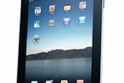
The iPad can be reliably used for preliminary assessment of lung nodules, offering accurate nodule detection and localization with only slightly longer image reading times than a conventional workstation, according to research presented at last week's European Congress of Radiology (ECR) in Vienna.
In a study comparing Apple's iPad 2 with an iMac desktop computer, a research team from the University of Pisa found that all nodules detected on the iMac were also seen and correctly localized on the iPad.
"Also, image reading was relatively fast [on the iPad] with no significant difference between the [iPad and iMac], which may support the hypothesis that the iPad 2 could be used reliably for preliminary visualization of lung nodules in this setting," said Dr. Lorenzo Faggioni. "Another potential additional application might be image sharing with nonradiology specialists and for teaching purposes."
Faggioni presented the results during a Monday scientific session at the congress.
Powerful mobile devices have become available that allow the display of a large amount of medical images from several imaging modalities, without the need for a dedicated standalone workstation, Faggioni said. One of those tablets is the popular iPad, and the researchers sought to evaluate the effectiveness of the iPad 2 as a mobile device for 2D reading of chest CT datasets for assessing pulmonary nodules.
The team retrospectively reviewed 28 chest CT exams with a total of 325 nodules sized between 2 mm and 34 mm. All CT images were acquired using a 64-detector-row CT scanner. The images were wirelessly imported in DICOM format from an Apple iMac desktop computer connected to the institution's hospital PACS to a 64-GB iPad 2 running the OsiriX HD open-source image viewing software.
Two experienced readers read the CT datasets independently, first on the 27-inch iMac (using the OsiriX 3.9 software) and then on the iPad 2. Detection rate and segmentation localization were recorded for each dataset, as well as the time needed to complete the reading of each chest CT study. The readers also rated image quality on a three-point scale, with 1 representing poor, 2 meaning fair, and 3 meaning good.
All nodules detected on the iMac were also identified on the iPad 2. In addition, nodule segmentation localization was correct in all cases, Faggioni said. Image quality was rated as good with both devices.
Image reading time was also comparable between the two devices, with an average of 4.72 ± 2.23 minutes for the iMac and 5.14 ± 2.56 minutes for the iPad 2 (p > 0.05).
"We found that the iPad 2 can be used successfully for 2D reading of chest CT datasets in patients with pulmonary nodules, as all lesions detected on the iMac were also found and correctly localized on the iPad 2," he said.


![Examples of ultrasound findings and techniques. (A) Images in a 39-year-old male patient with a mass in the left thigh. The mass is heterogeneous on the B-mode US image (compared with the patient in D) and showed increased microvascularity (superb microvascular imaging [SMI]) and shear-wave elastography (SWE) values. Undifferentiated pleomorphic sarcoma was diagnosed at biopsy (with pleomorphic rhabdomyosarcoma in surgical specimen). (B) Images in an 18-year-old male patient with a mass in the left leg. The mass is hypoechoic on the B-mode image, with no other findings suggestive of malignancy. The lesion is in contact with the cortex of the tibia, which is slightly irregular. CT revealed a doubtful anteromedial tibial erosion. The microvascular study demonstrated high vascularization, suggestive of malignancy. Periosteal Ewing sarcoma was diagnosed with both histologic and immunohistochemical confirmation. (C) Images in a 69-year-old female patient with a lump growing on the outside of the left leg. Multiple SWE examinations were performed (please note the high values obtained in the measurements, whereas the color map highlights the stiffness relative to adjacent tissues). SMI showed areas of increased vascularization to target for sampling. Undifferentiated spindle cell sarcoma was diagnosed at biopsy, with residual leiomyosarcoma in the surgical specimen after neoadjuvant therapy. (D) Images in a 56-year-old female patient with a mass in the right thigh. The mass is heterogeneous at both B-mode ultrasound (similar to patient A) and MRI (coronal T2-weighted spectral attenuated inversion recovery [SPAIR]; T1-weighted pre-contrast and postcontrast imaging), which even shows uptake after the administration of paramagnetic contrast material, which is traditionally suggestive of malignancy. Low values at SMI and elastography are suggestive of benignity. Spindle cell lipoma was diagnosed at biopsy, with atypical spindle cell lipomatous tumor in the surgical specimen.](https://img.auntminnieeurope.com/mindful/smg/workspaces/default/uploads/2026/08/images-radiol250278fig2.APCFLSvX6p.jpg?auto=format%2Ccompress&fit=crop&h=100&q=70&w=100)


![Examples of ultrasound findings and techniques. (A) Images in a 39-year-old male patient with a mass in the left thigh. The mass is heterogeneous on the B-mode US image (compared with the patient in D) and showed increased microvascularity (superb microvascular imaging [SMI]) and shear-wave elastography (SWE) values. Undifferentiated pleomorphic sarcoma was diagnosed at biopsy (with pleomorphic rhabdomyosarcoma in surgical specimen). (B) Images in an 18-year-old male patient with a mass in the left leg. The mass is hypoechoic on the B-mode image, with no other findings suggestive of malignancy. The lesion is in contact with the cortex of the tibia, which is slightly irregular. CT revealed a doubtful anteromedial tibial erosion. The microvascular study demonstrated high vascularization, suggestive of malignancy. Periosteal Ewing sarcoma was diagnosed with both histologic and immunohistochemical confirmation. (C) Images in a 69-year-old female patient with a lump growing on the outside of the left leg. Multiple SWE examinations were performed (please note the high values obtained in the measurements, whereas the color map highlights the stiffness relative to adjacent tissues). SMI showed areas of increased vascularization to target for sampling. Undifferentiated spindle cell sarcoma was diagnosed at biopsy, with residual leiomyosarcoma in the surgical specimen after neoadjuvant therapy. (D) Images in a 56-year-old female patient with a mass in the right thigh. The mass is heterogeneous at both B-mode ultrasound (similar to patient A) and MRI (coronal T2-weighted spectral attenuated inversion recovery [SPAIR]; T1-weighted pre-contrast and postcontrast imaging), which even shows uptake after the administration of paramagnetic contrast material, which is traditionally suggestive of malignancy. Low values at SMI and elastography are suggestive of benignity. Spindle cell lipoma was diagnosed at biopsy, with atypical spindle cell lipomatous tumor in the surgical specimen.](https://img.auntminnieeurope.com/mindful/smg/workspaces/default/uploads/2026/08/images-radiol250278fig2.APCFLSvX6p.jpg?auto=format%2Ccompress&fit=crop&h=112&q=70&w=112)