CHICAGO - Researchers from a top London hospital have given a resounding vote of confidence to the clinical value of digital breast tomosynthesis (DBT), after assessing its potential in cancer screening and diagnosis in more than 1,500 women during the past two years.
DBT is more accurate than 2D mammography and film-screen mammography in the interpretation of abnormalities, can predict histological tumor size by clearly demonstrating the margins and extent of mammographic lesions, and is more accurate than supplementary magnification views in the diagnostic workup of soft mammographic lesions, according to Dr. Margaret Adejolu, a radiologist from King's College Hospital.
"DBT is an application of digital mammography that allows 3D evaluation of the breast. Its value lies in its ability to reduce or eliminate the effects of overlapping tissue, a drawback of conventional 2D mammography which significantly reduces its sensitivity in detecting breast lesions and causes false-positive results," she wrote in an RSNA education exhibit.
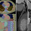
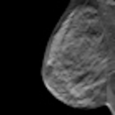
At King's College, researchers use the Hologic Selenia Dimensions system, which can perform 2D mammography, DBT, or both in the same compression. The DBT examination involves the acquisition of images of the stationary compressed breast at multiple angles. The x-ray tube moves through an arc of 15°, and a series of low-dose exposures are made during the tube movement. Data from the exposures is then used to reconstruct 1-mm slices for viewing in a plane parallel to the digital detector. Reconstructed images can be viewed by scrolling through the slices individually or in a dynamic cine mode.
"DBT helps to confirm or exclude the presence of genuine mammographic abnormalities when 2D mammography is equivocal. Focal asymmetries and architectural distortions constitute the majority of equivocal mammographic abnormalities," Adejolu noted.
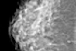
Malignancy presenting as a focal asymmetry will usually reveal an additional mammographic abnormality such as a mass or distortion on DBT. Therefore, DBT can improve diagnostic certainty in attributing a focal asymmetry to asymmetric glandular tissue. Genuine architectural distortions that appear equivocal on 2D mammography usually show a dramatic improvement in the conspicuity of the lesion on DBT, she stated.
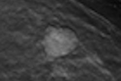
DBT can improve lesion characterization and allow for more accurate BI-RADS classification by providing better visualization of the margins of breast lesions. It can demonstrate an additional mammographic abnormality that is more representative of the underlying pathology. For instance, a BI-RADS 5 tissue lesion that is occult on 2D mammography may be shown in association with BI-RADS 3 microcalcification.
Furthermore, DBT may help to identify additional tumor foci that are not readily evident on 2D mammography, Adejolu continued. About 30% of women with breast cancer have multifocal or multicentric tumors, and invasive lobular cancers have a disproportionately higher incidence of multiple breast cancers. Dynamic contrast-enhanced MRI is the gold standard for excluding additional tumor foci, but many centers routinely perform whole-breast ultrasound as an adjunct to 2D mammography for this purpose and reserve breast MR for patients at highest risk of developing multiple cancers. DBT is a potentially cheaper alternative to MRI, but its sensitivity for detecting additional lesions compared with MR must be determined by further studies.
"We have found that DBT delineates the margins and extent of a lesion, and thus the histological size of breast lesions, more accurately than 2D mammography, providing vital information for prospective surgical planning," Adejolu noted.
The 3D location of a breast lesion can be accurately determined from its position within a DBT slice. It can provide an accurate road map for second-look targeted ultrasound when small lesions are identified on 2D mammography and are not readily identified on ultrasound, and can provide guidance for localization when this is not possible using ultrasound guidance. Also, the technique can confirm the intradermal location of suspicious microcalcification when it is found within the most peripheral DBT slab on the given view.
Overall, DBT is effective in the diagnostic workup and management of breast lesions, but further large-scale studies are now being conducted to evaluate the possible role of DBT in breast screening in Europe, Adejolu concluded.