The development of contrast-enhanced ultrasound (CEUS) quantification studies holds great promise in the gastrointestinal (GI) tract and is an exciting future possibility, according to a leading expert from Italy.
"We are recruiting patients and collecting the data. They are interesting in some cases but much work remains to be done to determine the definitive role of these new quantitative techniques in clinical practice," Dr. Carla Serra from the department of GI disease and internal medicine at S. Orsola Malpighi Hospital, University of Bologna, told delegates at the recent World Federation for Ultrasound in Medicine and Biology (WFUMB) congress in Vienna.
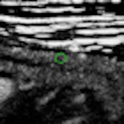
 Contrast-enhanced ultrasound of the bowel after sulphur hexafluoride-filled microbubble injection. Analysis of bowel wall perfusion with a quantitative software (intensity curve versus time). Image courtesy of Dr. Carla Serra.
CEUS is already showing potential in chronic inflammatory bowel disease evaluation, especially for the assessment of activity in Crohn's disease (CD), she noted. The technique involves intravenous administration of an ultrasound contrast agent with real-time examination, and can provide an accurate depiction of the bowel wall microvascularization and the perienteric tissues. The introduction of quantification techniques enables objective quantitative measurement of the enhancement.
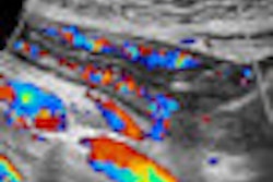
"With color power Doppler, we can see macro- and microvessels in the intestinal wall, and studies show that it can evaluate CD activity before and after therapy," Serra said.
To obtain an adequate quantitative imaging analysis, it is mandatory that the modality of acquisition of the contrast-enhanced clips is standardized to reduce variability not linked to the effect of the treatment. Semiquantitative methods can identify different patterns of enhancement or measure the entire wall and enhancement portion, but these techniques lack standardization.
"This is the great problem with ultrasound," she explained. "But the solution could be to use quantitative methods. New software with standardized procedures provides an objective evaluation of the amount of contrast and is coming in clinical practice."
|
Typical problems encountered with contrast-enhanced ultrasound
Source: Dr. Carla Serra, University of Bologna, WFUMB 2011 |
The software makes it feasible to immediately export data in Excel and collect it for future patient visits or comparison with other patients. In a quantification method for CD, it is possible to view the region-of-interest and, during the injection time, the arrival and amount of contrast, as well as the typical wash-in and wash-out curves. Several studies have shown that this technique has both high specificity and sensitivity in evaluating treatment results, but translating these advances into clinical practice remains challenging because one examination is still very different to another.
"The crucial point is to reduce the variability not linked to the treatment effect," Serra said. "There are many problems, starting with the technique itself. CEUS is a very difficult tool that requires a standardized method. We must use the same probes, positions, loops, and presets, otherwise it is very hard to compare patients or observe the same patient at a later stage."
Another challenge is the gut target. In CD cases, it is essential to determine the best portion of bowel to evaluate, and loops at different depths and gut movements also complicate the ultrasound examination. Also, problems related to patient cardiac output or medication may influence the investigation, and difficulties with contrast injection procedure or scanning settings may occur.
"We must be sure to have basic research to validate parameters in clinical practice. We need intraobserver stability, stability toward external factors, reproducibility, and then we can compare different software," she said.
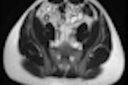
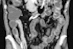
Overall, though, CEUS appears to be about as accurate as CT and MRI for detecting intramural and extramural extension in CD patients. B-mode ultrasound can evaluate the localization and length of the affected intestinal segments and allow identification of transmural complications, stenosis, and intestinal obstruction. Furthermore, Doppler techniques are proving useful for visualizing and quantifying bowel vascularization.