A new iterative reconstruction program permits the acquisition of high-quality body CT angiography (CTA) images using about half the dose of filtered back projection (FBP) imaging, a study in European Radiology concludes.
At 50% dose reduction, raw-data-based iterative reconstruction with the sinogram-affirmed iterative reconstruction (SAFIRE) technique actually demonstrated less image noise than the full-dose FBP images, while sharpening delineation of the aorta, according to researchers from Switzerland.
"Preliminary evidence suggests that raw data-based iterative reconstructions have the potential to reduce radiation dose by more than 50% in body CTA studies without deterioration in image quality," wrote Dr. Anna Winklehner and colleagues from the Institute of Diagnostic and Interventional Radiology at University Hospital Zurich.
Tube current reduction has traditionally been the method of choice for CT dose reduction, but the drawback is greatly increased image noise, especially with the use of FBP reconstruction, they wrote (European Radiology, 6 August 2011).
Several studies have shown the benefit of domain-based iterative reconstruction techniques in clinical applications in the chest and abdomen, with image quality improvements that can be used to reduce the radiation dose. Among the newest iterative reconstruction methods is SAFIRE. But rather than the domain-based techniques used in earlier techniques, SAFIRE relies on a noise modeling technique supported by the raw image data (sinogram data) that reduces noise and maintains image sharpness.
"SAFIRE estimates the local noise content in each image pixel by analyzing the raw data contributing to this pixel, and removes it from the current image data set," Winklehner et al wrote. "This is done step-by-step with up to five iterations. Each iteration leads to further noise reduction."
The study aimed to prospectively evaluate the dose-saving potential of the SAFIRE method compared to FBP in patients undergoing body CTA.
The group examined 25 patients using thoraco-abdominal CTA on a 128-slice dual-source CT (SOMATOM Definition Flash, Siemens Healthcare) following administration of 80 mL iodinated contrast and a 40-mL saline flush. The effective tube current was 164-401 mAs using attenuation-based tube current modulation (CAREDose4D), slice acquisition 2×64×0.6 mm, gantry rotation time 500 ms, and pitch 1. Both tubes were operated at 120 kV.
Filtered back projection (FBP) was applied to the full dose images obtained using data from both x-ray tubes, while half-dose datasets were reconstructed from only one tube-detector system, effectively halving the dose. The SAFIRE technique (set at strength 3 from a range of 1-5) was applied to the half-dose images and FBP was applied to the full-dose (two tube) images.
The authors assessed image quality and sharpness of the aortic contour, measured vessel attenuation and noise, and calculated the contrast-to-noise ratio. Factors that diminished overall image quality such as noise, metal artifacts, motion, and contrast-related artifacts were assessed on a four-point scale, with one being optimum and four severe, meaning that image quality was affected.
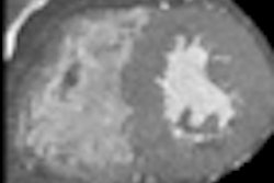
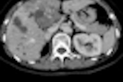
 Images of the upper abdomen reconstructed using filtered backprojection (FBP) and the sinogram affirmed iterative reconstruction (SAFIRE) algorithm. Above, transverse image reconstruction with standard FBP at the level of the upper abdomen shows a high level of image noise. All images courtesy of Dr. Alkadhi Hatem.
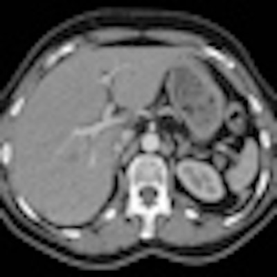
 Image reconstruction with raw data-based SAFIRE shows markedly reduced image noise and sharper depiction of vessel contours.
Half the dose with far less noise
Noise that deteriorated image quality occurred in 24/25 (96%) of the half-dose FBP images but not in full-dose FBP and HD-raw databased iterative reconstruction datasets (p < 0.001), the authors reported. Other artifacts occurred with similar prevalence among the datasets, they said, and no significant differences were found between the three image datasets for the presence or absence of other artifacts besides noise (p > 0.05). The inter-reader agreement for image quality analysis was moderate to good (kappa = 0.579 - 0.701).
Finally, sharpness of the aortic contour was found to be significantly better for the full-dose FBP (median score 1) and half-dose SAFIRE (median score 1) versus the half-dose FBP images (median score 2, p < 0.001).
The estimated effective dose was 21.7± 5.8 mSv for the full-dose (two x-ray tubes) CT exams, and 10.9±2.9 mSv (effective dose/2) for the half-dose examinations.
The comparison of image datasets in patients undergoing CTA showed that raw data-based SAFIRE can potentially reduce doses more than 50% versus standard reconstruction with FBP while maintaining diagnostic image quality, Winklehner et al wrote.
"Conventional CT image reconstruction approaches like back-projection type algorithms comprise a trade-off between sharpness and noise," the authors explained. "Sharpness, i.e. the minimum visible detail size, can only be increased at the expense of higher image noise, or vice versa, noise can only be reduced by lowering the sharpness. This trade-off limits the minimum radiation dose required for a specific diagnostic application. SAFIRE represents an iterative optimization process which overcomes this constraint."
SAFIRE reconstruction uses a noise modeling technique, based on raw data that utilizes the "known propagation of noise in projection data" into the image domain, they wrote. Reconstructed image data consists of the raw data plus the image noise. "The goal is to separate the information and noise with a maximum likelihood given by the model," Winklehner et al wrote. "This task can be translated into a mathematical optimization problem and solved iteratively." In SAFIRE the noise content is estimated and subtracted from the current dataset in each of five iteration loops.
The authors cited study limitations including the small patient population, the lack of patients having a body mass index (BMI) greater than 35, and the lack of diagnostic performance assessment -- only image-quality evaluation -- among the readers.
Noise was significantly lower in the half-dose datasets using SAFIRE, suggesting that dose reduction could be carried much further without reducing image quality, Winklehner and colleagues wrote.