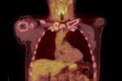
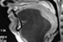
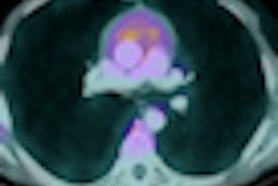
Ultrasound is a complementary procedure to PET/CT for diagnosing recurring head and neck cancer, according to an article in press published on 22 July in the Reports on Practical Oncology and Radiotherapy. Identifying recurring cancer early enough can mean the difference between good odds of survival versus a more rapid demise.
Researchers at the Greater Poland Medical Centre in Poznan conducted a retrospective study of patients treated for head and neck squamous cell carcinoma to compare the diagnostic performance of PET/CT and ultrasound, as well as their combined use to detect local recurrence. Their objective was to determine the effectiveness of the examinations with respect to identifying cancer in patients whose clinical otolaryngological exams did not reveal any sign of cancer recurrence.
The study included 83 patients treated between December 2007 and April 2010. The majority of patients (43%) had cancer of the oral cavity and almost one-fourth (24%) had cancer of the larynx. The remainder had cancer in their nasal cavity and paranasal sinuses (13%), salivary gland (8%), hypopharynx (7%), and oropharynx (5%).
The patients underwent regularly scheduled examinations following their treatment. They underwent both an ultrasound exam and a PET/CT exam, with a mean of 14 months following treatment.
A total of 36 patients developed a recurrence at a mean follow-up of 10 months, none of whom exhibited any clinical indications of cancer. PET/CT had sensitivity of 86% and specificity 82%, and ultrasound had sensitivity of 81% and specificity of 87%.
The positive predictive value was 83% for ultrasound and 79% for PET/CT. Negative predictive value was slightly higher for PET/CT at 89% compared with 85% for ultrasound.
Lead author Dr. Malgorzata Wierzbicka, of the department of otolaryngology of Poznan University of Medical Sciences, and colleagues relied on lymph node size and shape criteria to determine abnormalities, whereas PET/CT provided additional information about metabolism and precise localization of the occult recurrence.
"Ultrasound offers numerous advantages over PET/CT," the authors wrote. "It is less time-consuming, inexpensive, repeatable, well-tolerated by patients, and does not expose a patient to radiation." They noted that its disadvantages were interobserver variability and the fact that imaging was limited to soft tissue.
The value of PET/CT is greater in ruling out recurrence, according to the authors. "PET/CT alone cannot reliably predict the presence of residual cervical metastatic disease, but its negative predictive value is high enough to rule recurrence out. A negative result can reliably allow the patient to avoid neck dissection and further images."
When recurring head and neck cancers are diagnosed early, a patient with an early-stage tumor who undergoes salvage surgery has a 70% rate of two-year relapse-free survival, compared with a 22% rate for a patient with an advanced stage tumor.


![Examples of ultrasound findings and techniques. (A) Images in a 39-year-old male patient with a mass in the left thigh. The mass is heterogeneous on the B-mode US image (compared with the patient in D) and showed increased microvascularity (superb microvascular imaging [SMI]) and shear-wave elastography (SWE) values. Undifferentiated pleomorphic sarcoma was diagnosed at biopsy (with pleomorphic rhabdomyosarcoma in surgical specimen). (B) Images in an 18-year-old male patient with a mass in the left leg. The mass is hypoechoic on the B-mode image, with no other findings suggestive of malignancy. The lesion is in contact with the cortex of the tibia, which is slightly irregular. CT revealed a doubtful anteromedial tibial erosion. The microvascular study demonstrated high vascularization, suggestive of malignancy. Periosteal Ewing sarcoma was diagnosed with both histologic and immunohistochemical confirmation. (C) Images in a 69-year-old female patient with a lump growing on the outside of the left leg. Multiple SWE examinations were performed (please note the high values obtained in the measurements, whereas the color map highlights the stiffness relative to adjacent tissues). SMI showed areas of increased vascularization to target for sampling. Undifferentiated spindle cell sarcoma was diagnosed at biopsy, with residual leiomyosarcoma in the surgical specimen after neoadjuvant therapy. (D) Images in a 56-year-old female patient with a mass in the right thigh. The mass is heterogeneous at both B-mode ultrasound (similar to patient A) and MRI (coronal T2-weighted spectral attenuated inversion recovery [SPAIR]; T1-weighted pre-contrast and postcontrast imaging), which even shows uptake after the administration of paramagnetic contrast material, which is traditionally suggestive of malignancy. Low values at SMI and elastography are suggestive of benignity. Spindle cell lipoma was diagnosed at biopsy, with atypical spindle cell lipomatous tumor in the surgical specimen.](https://img.auntminnieeurope.com/mindful/smg/workspaces/default/uploads/2026/08/images-radiol250278fig2.APCFLSvX6p.jpg?auto=format%2Ccompress&fit=crop&h=100&q=70&w=100)


![Examples of ultrasound findings and techniques. (A) Images in a 39-year-old male patient with a mass in the left thigh. The mass is heterogeneous on the B-mode US image (compared with the patient in D) and showed increased microvascularity (superb microvascular imaging [SMI]) and shear-wave elastography (SWE) values. Undifferentiated pleomorphic sarcoma was diagnosed at biopsy (with pleomorphic rhabdomyosarcoma in surgical specimen). (B) Images in an 18-year-old male patient with a mass in the left leg. The mass is hypoechoic on the B-mode image, with no other findings suggestive of malignancy. The lesion is in contact with the cortex of the tibia, which is slightly irregular. CT revealed a doubtful anteromedial tibial erosion. The microvascular study demonstrated high vascularization, suggestive of malignancy. Periosteal Ewing sarcoma was diagnosed with both histologic and immunohistochemical confirmation. (C) Images in a 69-year-old female patient with a lump growing on the outside of the left leg. Multiple SWE examinations were performed (please note the high values obtained in the measurements, whereas the color map highlights the stiffness relative to adjacent tissues). SMI showed areas of increased vascularization to target for sampling. Undifferentiated spindle cell sarcoma was diagnosed at biopsy, with residual leiomyosarcoma in the surgical specimen after neoadjuvant therapy. (D) Images in a 56-year-old female patient with a mass in the right thigh. The mass is heterogeneous at both B-mode ultrasound (similar to patient A) and MRI (coronal T2-weighted spectral attenuated inversion recovery [SPAIR]; T1-weighted pre-contrast and postcontrast imaging), which even shows uptake after the administration of paramagnetic contrast material, which is traditionally suggestive of malignancy. Low values at SMI and elastography are suggestive of benignity. Spindle cell lipoma was diagnosed at biopsy, with atypical spindle cell lipomatous tumor in the surgical specimen.](https://img.auntminnieeurope.com/mindful/smg/workspaces/default/uploads/2026/08/images-radiol250278fig2.APCFLSvX6p.jpg?auto=format%2Ccompress&fit=crop&h=112&q=70&w=112)