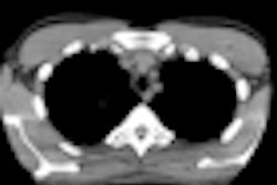
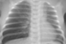
In a typical tertiary care hospital in Germany, only 30% of pediatric imaging exams performed emit ionizing radiation. For these procedures, however, radiation dose exposure needs to be kept low due to the patients' highly radiosensitive tissues, according to a new study.
Two pediatric radiologists describe the most effective methods of lowering the exposure of children to ionizing radiation without sacrificing diagnostic reliability in an article published in the 17 June issue of Deutsches Ärzeblatt International (2011, Vol. 108:24, pp. 407-414). Their focus combines time-honored shielding recommendations with utilization of features in the newest generations of imaging modality equipment.
The best radiation protection for pediatric patients are physicians with optimum training in ultrasound, noted lead author Dr. Gerhard Alzen, chair of pediatric radiology at University Hospital Giessen. A physician experienced in interpreting ultrasound exams may be able to answer questions using just this modality, or may be able to better order the most appropriate exam when ultrasound images do not provide a diagnosis.
Alzen and co-author Dr. Gabriele Benz-Bohm, of the University of Cologne's division of pediatric radiology, emphasize that radiologists need to corroborate with referring physicians and discuss clinical indications. When the diagnostic question is clear, radiologists and radiographers can recommend and perform the most appropriate exam for an individual patient, at the lowest possible radiation dose without compromising diagnostic quality. The authors also recommend that radiographers be proficient in knowing operating procedures, guidelines, and dose limits in view of the complexity of age- and indication-dependent machine adjustment for pediatric examinations.
Alzen and Benz-Bohm describe features of radiography units that are child-friendly with respect to optimizing dose for pediatric exams, and should be included when purchasing new equipment. Their guidelines also discuss the proper setting of tube voltage, the use of tube filters, the limited use of scattered-radiation grids, and suitable patient positioning. They recommend the compulsory infant holders and other fixation devices.
Pediatric CT exam tips include the use of an image gallery, because availability of reference images has proved to be effective in reducing radiation dose. By using an image gallery, the University Hospital RWTH Aachen in Germany achieved a dose reduction of more than 60% for abdominal and pelvic examinations. The authors suggest that an image gallery containing example scans of patients at different ages enables CT examination parameters to be adjusted to individual ages, based on specific information needed from the CT scan for each CT scanner being used. They recommend that example images acquired from the use of simulation also show different doses.
In addition, Alzen and Benz-Bohm recommend that radiology departments distribute the patient x-ray exam record card that is available on the Image Gently website. The cards are a record of cumulative radiation dose and are a tangible way to raise awareness of cumulative radiation dose risk, they noted.


![Examples of ultrasound findings and techniques. (A) Images in a 39-year-old male patient with a mass in the left thigh. The mass is heterogeneous on the B-mode US image (compared with the patient in D) and showed increased microvascularity (superb microvascular imaging [SMI]) and shear-wave elastography (SWE) values. Undifferentiated pleomorphic sarcoma was diagnosed at biopsy (with pleomorphic rhabdomyosarcoma in surgical specimen). (B) Images in an 18-year-old male patient with a mass in the left leg. The mass is hypoechoic on the B-mode image, with no other findings suggestive of malignancy. The lesion is in contact with the cortex of the tibia, which is slightly irregular. CT revealed a doubtful anteromedial tibial erosion. The microvascular study demonstrated high vascularization, suggestive of malignancy. Periosteal Ewing sarcoma was diagnosed with both histologic and immunohistochemical confirmation. (C) Images in a 69-year-old female patient with a lump growing on the outside of the left leg. Multiple SWE examinations were performed (please note the high values obtained in the measurements, whereas the color map highlights the stiffness relative to adjacent tissues). SMI showed areas of increased vascularization to target for sampling. Undifferentiated spindle cell sarcoma was diagnosed at biopsy, with residual leiomyosarcoma in the surgical specimen after neoadjuvant therapy. (D) Images in a 56-year-old female patient with a mass in the right thigh. The mass is heterogeneous at both B-mode ultrasound (similar to patient A) and MRI (coronal T2-weighted spectral attenuated inversion recovery [SPAIR]; T1-weighted pre-contrast and postcontrast imaging), which even shows uptake after the administration of paramagnetic contrast material, which is traditionally suggestive of malignancy. Low values at SMI and elastography are suggestive of benignity. Spindle cell lipoma was diagnosed at biopsy, with atypical spindle cell lipomatous tumor in the surgical specimen.](https://img.auntminnieeurope.com/mindful/smg/workspaces/default/uploads/2026/08/images-radiol250278fig2.APCFLSvX6p.jpg?auto=format%2Ccompress&fit=crop&h=100&q=70&w=100)


![Examples of ultrasound findings and techniques. (A) Images in a 39-year-old male patient with a mass in the left thigh. The mass is heterogeneous on the B-mode US image (compared with the patient in D) and showed increased microvascularity (superb microvascular imaging [SMI]) and shear-wave elastography (SWE) values. Undifferentiated pleomorphic sarcoma was diagnosed at biopsy (with pleomorphic rhabdomyosarcoma in surgical specimen). (B) Images in an 18-year-old male patient with a mass in the left leg. The mass is hypoechoic on the B-mode image, with no other findings suggestive of malignancy. The lesion is in contact with the cortex of the tibia, which is slightly irregular. CT revealed a doubtful anteromedial tibial erosion. The microvascular study demonstrated high vascularization, suggestive of malignancy. Periosteal Ewing sarcoma was diagnosed with both histologic and immunohistochemical confirmation. (C) Images in a 69-year-old female patient with a lump growing on the outside of the left leg. Multiple SWE examinations were performed (please note the high values obtained in the measurements, whereas the color map highlights the stiffness relative to adjacent tissues). SMI showed areas of increased vascularization to target for sampling. Undifferentiated spindle cell sarcoma was diagnosed at biopsy, with residual leiomyosarcoma in the surgical specimen after neoadjuvant therapy. (D) Images in a 56-year-old female patient with a mass in the right thigh. The mass is heterogeneous at both B-mode ultrasound (similar to patient A) and MRI (coronal T2-weighted spectral attenuated inversion recovery [SPAIR]; T1-weighted pre-contrast and postcontrast imaging), which even shows uptake after the administration of paramagnetic contrast material, which is traditionally suggestive of malignancy. Low values at SMI and elastography are suggestive of benignity. Spindle cell lipoma was diagnosed at biopsy, with atypical spindle cell lipomatous tumor in the surgical specimen.](https://img.auntminnieeurope.com/mindful/smg/workspaces/default/uploads/2026/08/images-radiol250278fig2.APCFLSvX6p.jpg?auto=format%2Ccompress&fit=crop&h=112&q=70&w=112)