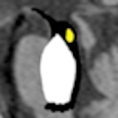
The sudden appearance of animals in radiology images is not the result of eye fatigue, but may be an easily recognizable, and highly memorable, sign of disease to be reported. Identifying the eye of a tiger, the face of a giant panda, a butterfly, or a penguin not only helps to narrow down the elaborate list of differentials, but can also facilitate the diagnosis on the basis of imaging findings alone, according to a study presented at the 2011 European Congress of Radiology (ECR) in Vienna.
"Animals in the brain" proved to be the second most popular poster with radiologists using the Electronic Presentation Online System (EPOS) during ECR. The authors, Drs. Ankur Arora and Abhay Kapoor from the department of radiodiagnosis at the Govind Ballabh Pant Hospital in New Delhi, India, received a Certificate of Merit from the judges of the Scientific Exhibition.
Based on a shared experience among radiologists that certain neurologic conditions have characteristic radiologic manifestations that resemble various types of animals, the authors gathered a rich glossary of these signs to assist colleagues. Keyword references to birds, insects, and the different body parts of animals illustrate classic neuroradiologic manifestations.
 The "penguin sign" may signal progressive supranuclear palsy. In this image, atrophy of the tectum, particularly the superior colliculus, can be seen. Image courtesy of Dr. Laughlin Dawes, Sir Charles Gairdner Hospital, Perth, Australia.
An example of these useful signs is the "penguin" seen in an MRI of the brain. More than amusing, the penguin is an interesting radiological sign seen in patients with progressive supranuclear palsy (PSP), noted Arora. It refers to atrophy of the midbrain tegmentum, with a relatively preserved pons on midsagittal T1-weighted images. The "penguin sign" can be helpful in distinguishing progressive supranuclear palsy from multisystem atrophy and Parkinson's disease.
In their poster, the authors provide 20 examples of animal-shaped radiological patterns and the conditions they may signal. "Face of the Giant Panda" is an uncommon intracranial manifestation of Wilson's disease caused by a combination of signal intensity changes at the level of midbrain on T2-weighted MR images. A "tiger-striped" cerebellar foliar pattern that consists of alternating bands on T1- and T2-weighted images is considered almost pathognomonic of Lhermitte-Duclos disease.
Beyond the animal kingdom, the authors are avid collectors of other patterns in radiology readings. In a letter to the Korean Journal of Radiology (November-December 2010, Vol. 11:6, pp. 702-703), they congratulated Dr. Dong Zhang, for his contribution of what they called a unique and interesting finding. He first identified the presence of a "notch sign" in neuro MRI readings that may help a radiologist in narrowing down the differentials in patients with suspected central nervous system lymphoma.







![Examples of ultrasound findings and techniques. (A) Images in a 39-year-old male patient with a mass in the left thigh. The mass is heterogeneous on the B-mode US image (compared with the patient in D) and showed increased microvascularity (superb microvascular imaging [SMI]) and shear-wave elastography (SWE) values. Undifferentiated pleomorphic sarcoma was diagnosed at biopsy (with pleomorphic rhabdomyosarcoma in surgical specimen). (B) Images in an 18-year-old male patient with a mass in the left leg. The mass is hypoechoic on the B-mode image, with no other findings suggestive of malignancy. The lesion is in contact with the cortex of the tibia, which is slightly irregular. CT revealed a doubtful anteromedial tibial erosion. The microvascular study demonstrated high vascularization, suggestive of malignancy. Periosteal Ewing sarcoma was diagnosed with both histologic and immunohistochemical confirmation. (C) Images in a 69-year-old female patient with a lump growing on the outside of the left leg. Multiple SWE examinations were performed (please note the high values obtained in the measurements, whereas the color map highlights the stiffness relative to adjacent tissues). SMI showed areas of increased vascularization to target for sampling. Undifferentiated spindle cell sarcoma was diagnosed at biopsy, with residual leiomyosarcoma in the surgical specimen after neoadjuvant therapy. (D) Images in a 56-year-old female patient with a mass in the right thigh. The mass is heterogeneous at both B-mode ultrasound (similar to patient A) and MRI (coronal T2-weighted spectral attenuated inversion recovery [SPAIR]; T1-weighted pre-contrast and postcontrast imaging), which even shows uptake after the administration of paramagnetic contrast material, which is traditionally suggestive of malignancy. Low values at SMI and elastography are suggestive of benignity. Spindle cell lipoma was diagnosed at biopsy, with atypical spindle cell lipomatous tumor in the surgical specimen.](https://img.auntminnieeurope.com/mindful/smg/workspaces/default/uploads/2026/08/images-radiol250278fig2.APCFLSvX6p.jpg?auto=format%2Ccompress&dpr=2&fit=crop&h=167&q=70&w=250)

