MRI is essential for initial diagnosis and follow-up of acute musculotendinous injuries, and it can enable clinicians to estimate precisely when an athlete can return to competition as well as the risk of recurrent injury, but evidence for its specific use remains limited and controversial, according to sports imaging experts.
Following these injuries -- including strain, contusion, and avulsion injuries -- MR images are often acquired to locate the lesion and assess its severity. High-quality imaging helps to plan and guide an athlete's rehabilitation, but a clinical evaluation must determine return-to-play decisions, wrote a multinational group of authors in an article published online on 16 September by Insights into Imaging.
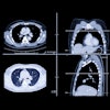
 A 24-year-old tennis player presented with left posterior thigh pain. Clinically, the lesion was diagnosed as a grade 2 injury. Axial fat-saturated T2-weighted turbo spin-echo image (A) and coronal short-tau inversion-recovery image (B) show a partial tear without retraction (grade 2 injury, yellow arrow) of the musculotendinous junction of the semimembranosus muscle. The tear measured 6 x 2 x 1.8 cm in size. S = short head of biceps femoris, L = long head of biceps femoris, M = semimembranosus; T = semitendinosus. All images courtesy of Dr. Daichi Hayashi, PhD.
"Fluid-sensitive MR sequences are suitable for detecting edematous changes in the muscles," noted lead author Dr. Daichi Hayashi, PhD, a research assistant professor of radiology at Boston University School of Medicine in the U.S. "T1-weighted sequences are used to differentiate between hemorrhage/hematoma and edema."
Furthermore, research efforts are ongoing to develop MRI techniques for assessing skeletal muscle ultrastructure. Diffusion tensor imaging (DTI) can provide detailed information on muscle structural changes, while muscle fiber tracking with DTI has been used successfully to assess muscle damage and measure pennation angle, he explained.
"Muscle is fairly uniform in bulk directionality and exhibits orderly arrangements on DTI. Following muscle injury, disturbance of the normal arrangement will be observed," Hayashi stated. "DTI and fiber-tracking packages are sold by all the major MR imaging vendors and can be performed using routinely available clinical scanners. Thus, DTI may become a useful tool in our clinical practice in the future."
Also, the use of MR spectroscopy (MRS) using phosphorus or sodium to assess skeletal muscle disorders has been explored. Phosphorus (31P) MRS measures oxidative metabolism, and sodium (23Na) MRS measures tissue sodium levels in muscle during exercise and recovery, he added. These techniques may be useful as a noninvasive tool in exercise physiology or sports medicine to better understand the dynamics of an exercising muscle, but their applicability and relevance to evaluation of muscle injury are yet to be determined.
 A 28-year-old man presented with right calf pain. Axial fat-saturated T2-weighted turbo spin-echo image demonstrate an area of hyperintensity with feathery appearance, consistent with a grade 1 injury (strain) of soleus muscle (yellow arrow). There is also epifascial hematoma and edema at the posterior aspects of the medial gastrocnemius (red arrows). SO = soleus; GM = medial head of gastrocnemius; GL = lateral head of gastrocnemius.
Over recent years, MRI has become a valuable tool for evaluation of traumatic muscle injuries, especially among football players, dancers, track and field athletes, and other competitive athletes. Under normal circumstances, images from only the affected leg are acquired using a surface coil, but the appropriate coil should be selected to obtain the desired field-of-view. Imaging of the contralateral leg is performed in exceptional cases only (e.g., bilateral injury).
"There are high demands on sports medicine physicians to quantify the prognosis and to predict when the athlete can return to training and competition. Imaging may assist in the prognostication of the healing process and in predicting the risk of recurrence, but the decision on return to play cannot be dependent on the imaging findings alone and must be balanced against the clinical situation," the authors stressed.
No official guidelines on the role of MRI or ultrasound in evaluating muscle injuries in high-level athletes have been published, and an algorithm that integrates clinical and imaging information into an explicit management plan would be very useful and needs to be developed, according to the authors.
"Ultrasound may play a role as an adjunct imaging method, but it is less accurate than MR imaging for assessing the extent of the injury and it cannot differentiate between new and old injuries," the authors concluded.