Dear AI Insider,
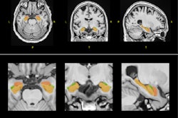
The development of viable and affordable AI technology remains frustratingly slow in some areas of clinical practice, but Alzheimer's disease identification is one field in which the software appears to be relatively advanced and there is good reason for optimism.
Today's top article provides a succinct and readable summary of how exactly AI can improve the care of Alzheimer's patients. Significantly, the authors won a prestigious magna cum laude award at ECR 2024 for their work.
Another recent study has shown that intelligent virtual and AI-based automated collimation functions can have a positive impact on the efficiency of x-ray image acquisitions, saving time for radiographers.
In other news, new evidence suggests two commercially available AI software applications can yield different volume estimations on stroke CT perfusion studies, but the differences aren't substantial enough to affect patient eligibility for thrombectomy.
By using AI, diagnostics can be made faster, better, and more effective, Prof. Philipp Paprottka from Munich, Germany, said in a Q&A interview. In interventional procedures, progress is being made through the further development of large-scale equipment and the improvement of navigation, fusion systems, and methods used for controlling the interventions.
In case you missed it earlier in May, Dr. Charlene Liew from Singapore outlined the current state of AI in radiology and where it is going in a keynote presentation ("AI in Radiology: The Past Informs the Future") at the International Society of Magnetic Resonance in Medicine meeting.
Philip Ward
Editor in Chief
AuntMinnieEurope.com





![A normal mammogram confirmed by three-year radiologic follow-up illustrates reader-marked regions of interest (ROIs) during (A) unaided (round 1) and (B) artificial intelligence (AI)–assisted (round 2) reading. Each colored dot represents an ROI for recall by a human reader. Readers could mark more than one ROI per case, represented by multiple dots of the same color. During AI-assisted reading, the AI system displayed three visible prompts: two with suspicion of malignancy scores of 35% (left mediolateral oblique [L MLO] and craniocaudal [L CC]) and one with a suspicion of malignancy score of 10% (right craniocaudal [R CC]), shown as polygonal overlays. Without AI, six of 10 readers (60%) marked a false-positive ROI. With AI assistance, this fell to two of 10 (20%). R MLO = right mediolateral oblique.](https://img.auntminnieeurope.com/mindful/smg/workspaces/default/uploads/2026/07/2026-07-14-radiology-mammogram-ai-auto-bias.H0bYO8QlWs.jpg?auto=format%2Ccompress&fit=crop&h=100&q=70&w=100)


![A normal mammogram confirmed by three-year radiologic follow-up illustrates reader-marked regions of interest (ROIs) during (A) unaided (round 1) and (B) artificial intelligence (AI)–assisted (round 2) reading. Each colored dot represents an ROI for recall by a human reader. Readers could mark more than one ROI per case, represented by multiple dots of the same color. During AI-assisted reading, the AI system displayed three visible prompts: two with suspicion of malignancy scores of 35% (left mediolateral oblique [L MLO] and craniocaudal [L CC]) and one with a suspicion of malignancy score of 10% (right craniocaudal [R CC]), shown as polygonal overlays. Without AI, six of 10 readers (60%) marked a false-positive ROI. With AI assistance, this fell to two of 10 (20%). R MLO = right mediolateral oblique.](https://img.auntminnieeurope.com/mindful/smg/workspaces/default/uploads/2026/07/2026-07-14-radiology-mammogram-ai-auto-bias.H0bYO8QlWs.jpg?auto=format%2Ccompress&fit=crop&h=112&q=70&w=112)