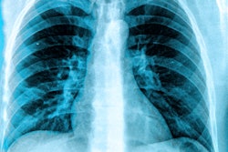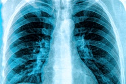
A new University College London Hospitals NHS Foundation Trust study aims to reduce the time taken to diagnose lung cancer by using Qure.ai's qXR technology to detect abnormalities on chest x-rays.
The LungIMPACT trial will investigate the use of this technology, which is designed to detect potential abnormalities on chest x-rays. It will be co-led by Nick Woznitza, PhD, and David Baldwin, MD.
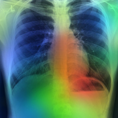
For the study, qXR will produce a secondary image for each chest x-ray with an overlay to highlight certain abnormalities. If the AI detects a problem, this will highlight the x-ray on the reporting worklist. This will allow for the reporting radiographer to prioritize this for urgent reporting. From there, the researchers will determine whether use of this AI "triage" can successfully bring down time diagnosis times.
This work was commissioned and funded by the NHS Cancer Programme, with the support of SBRI Healthcare and the NHS Accelerated Access Collaborative. It is supported by the National Institute for Health and Care Research Biomedical Research Centre at UCLH.




![A normal mammogram confirmed by three-year radiologic follow-up illustrates reader-marked regions of interest (ROIs) during (A) unaided (round 1) and (B) artificial intelligence (AI)–assisted (round 2) reading. Each colored dot represents an ROI for recall by a human reader. Readers could mark more than one ROI per case, represented by multiple dots of the same color. During AI-assisted reading, the AI system displayed three visible prompts: two with suspicion of malignancy scores of 35% (left mediolateral oblique [L MLO] and craniocaudal [L CC]) and one with a suspicion of malignancy score of 10% (right craniocaudal [R CC]), shown as polygonal overlays. Without AI, six of 10 readers (60%) marked a false-positive ROI. With AI assistance, this fell to two of 10 (20%). R MLO = right mediolateral oblique.](https://img.auntminnieeurope.com/mindful/smg/workspaces/default/uploads/2026/07/2026-07-14-radiology-mammogram-ai-auto-bias.H0bYO8QlWs.jpg?auto=format%2Ccompress&fit=crop&h=100&q=70&w=100)


![A normal mammogram confirmed by three-year radiologic follow-up illustrates reader-marked regions of interest (ROIs) during (A) unaided (round 1) and (B) artificial intelligence (AI)–assisted (round 2) reading. Each colored dot represents an ROI for recall by a human reader. Readers could mark more than one ROI per case, represented by multiple dots of the same color. During AI-assisted reading, the AI system displayed three visible prompts: two with suspicion of malignancy scores of 35% (left mediolateral oblique [L MLO] and craniocaudal [L CC]) and one with a suspicion of malignancy score of 10% (right craniocaudal [R CC]), shown as polygonal overlays. Without AI, six of 10 readers (60%) marked a false-positive ROI. With AI assistance, this fell to two of 10 (20%). R MLO = right mediolateral oblique.](https://img.auntminnieeurope.com/mindful/smg/workspaces/default/uploads/2026/07/2026-07-14-radiology-mammogram-ai-auto-bias.H0bYO8QlWs.jpg?auto=format%2Ccompress&fit=crop&h=112&q=70&w=112)