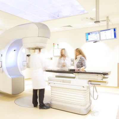
Varian, a Siemens Healthineers company, is showcasing its cancer care solutions, including advanced imaging, software, and services at the upcoming 2023 annual European Society for Radiotherapy and Oncology (ESTRO) meeting in Vienna, Austria.
The company is highlighting its HyperSight imaging software, Identify system, and Eclipse treatment planning system.
Hypersight uses conebeam CT that is faster than conventional linear accelerator-based imaging systems and can acquire large images, the company said. The software is available for the Halcyon and Ethos radiotherapy systems.
The Identify system addresses patient movement during radiotherapy, helping clinicians identify where the patient is during radiation treatment. The latest version of the system offers safety features such as automated beam hold functionality on the TrueBeam platform and process efficiency, the company added.
The latest version of the Eclipse system includes features that aim to improve accuracy in stereotactic radiosurgery treatments, as well as improve conformity for complex stereotactic body radiotherapy cases.
Finally, Varian said it is continuing to expand the capabilities of the Aria oncology information system, and it is introducing InSightive Gen2, a cloud-native analytics software.




![A normal mammogram confirmed by three-year radiologic follow-up illustrates reader-marked regions of interest (ROIs) during (A) unaided (round 1) and (B) artificial intelligence (AI)–assisted (round 2) reading. Each colored dot represents an ROI for recall by a human reader. Readers could mark more than one ROI per case, represented by multiple dots of the same color. During AI-assisted reading, the AI system displayed three visible prompts: two with suspicion of malignancy scores of 35% (left mediolateral oblique [L MLO] and craniocaudal [L CC]) and one with a suspicion of malignancy score of 10% (right craniocaudal [R CC]), shown as polygonal overlays. Without AI, six of 10 readers (60%) marked a false-positive ROI. With AI assistance, this fell to two of 10 (20%). R MLO = right mediolateral oblique.](https://img.auntminnieeurope.com/mindful/smg/workspaces/default/uploads/2026/07/2026-07-14-radiology-mammogram-ai-auto-bias.H0bYO8QlWs.jpg?auto=format%2Ccompress&fit=crop&h=100&q=70&w=100)


![A normal mammogram confirmed by three-year radiologic follow-up illustrates reader-marked regions of interest (ROIs) during (A) unaided (round 1) and (B) artificial intelligence (AI)–assisted (round 2) reading. Each colored dot represents an ROI for recall by a human reader. Readers could mark more than one ROI per case, represented by multiple dots of the same color. During AI-assisted reading, the AI system displayed three visible prompts: two with suspicion of malignancy scores of 35% (left mediolateral oblique [L MLO] and craniocaudal [L CC]) and one with a suspicion of malignancy score of 10% (right craniocaudal [R CC]), shown as polygonal overlays. Without AI, six of 10 readers (60%) marked a false-positive ROI. With AI assistance, this fell to two of 10 (20%). R MLO = right mediolateral oblique.](https://img.auntminnieeurope.com/mindful/smg/workspaces/default/uploads/2026/07/2026-07-14-radiology-mammogram-ai-auto-bias.H0bYO8QlWs.jpg?auto=format%2Ccompress&fit=crop&h=112&q=70&w=112)