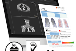
Advanced image analysis software developer QUIBIM has received the CE Mark for its Chest X-Ray Classification software application.
Available within QUIBIM's Precision image analysis platform, the artificial intelligence (AI)-based Chest X-Ray Classification tool is designed to help radiologists prioritize interpretation of radiographs that are potentially pathological, according to the company. The software is based on "referee" networks and convolutional neural networks that have been trained with a database of more than 500,000 images. After calculating the final probability of a chest x-ray being abnormal, the tool then utilizes these probabilities to estimate the presence of pathologies, QUIBIM said.



.png?auto=format%2Ccompress&fit=crop&h=167&q=70&w=250)


![A normal mammogram confirmed by three-year radiologic follow-up illustrates reader-marked regions of interest (ROIs) during (A) unaided (round 1) and (B) artificial intelligence (AI)–assisted (round 2) reading. Each colored dot represents an ROI for recall by a human reader. Readers could mark more than one ROI per case, represented by multiple dots of the same color. During AI-assisted reading, the AI system displayed three visible prompts: two with suspicion of malignancy scores of 35% (left mediolateral oblique [L MLO] and craniocaudal [L CC]) and one with a suspicion of malignancy score of 10% (right craniocaudal [R CC]), shown as polygonal overlays. Without AI, six of 10 readers (60%) marked a false-positive ROI. With AI assistance, this fell to two of 10 (20%). R MLO = right mediolateral oblique.](https://img.auntminnieeurope.com/mindful/smg/workspaces/default/uploads/2026/07/2026-07-14-radiology-mammogram-ai-auto-bias.H0bYO8QlWs.jpg?auto=format%2Ccompress&dpr=2&fit=crop&h=167&q=70&w=250)


