Dear Advanced Visualization Insider,
While multiparametric MRI can provide valuable morphological and functional information for diagnosing prostate cancer, these studies can be challenging for radiologists to interpret due to the various pulse sequences involved. Computer-aided detection (CAD) software can help, however, according to an Italian research team.
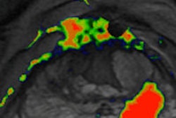
In a study presented at ECR 2014, Dr. Daniele Regge of the Institute for Cancer Research and Treatment in Candiolo shared the institution's experience with a CAD algorithm they developed that yields a color-coded map with per-pixel estimates of cancer probability. Click here to learn how well the system performed.
In other coverage from ECR 2014 in your Advanced Visualization Digital Community, AuntMinnieEurope.com Editor-in-Chief Philip Ward spoke in Vienna with mobile devices expert Dr. Erik Ranschaert in a video interview. Among other topics, Ranschaert discussed his recent survey of more than 200 radiologists on their use of tablets; more than half of radiologists reported that they currently utilized tablets in their clinical practice.
The use of mobile devices in radiology applications does require awareness of a number of crucial technical factors, however, according to a presentation by Rachel Toomey of the University College Dublin. Click here for all the details.
Stay tuned for additional coverage of advanced visualization topics from ECR 2014 in the coming weeks.
In other advanced visualization news, Italian researchers recently found that CT postprocessing software can play a key role in analyzing gunshot injuries. Their study indicates that 2D multiplanar reformatting and 3D volume rendering were shown to provide valuable additional information to axial imaging, particularly in examining vessels and bone lesions. Get all the details here.
Do you have an idea for a topic you'd like to see covered? As always, please feel free to email me.

![A normal mammogram confirmed by three-year radiologic follow-up illustrates reader-marked regions of interest (ROIs) during (A) unaided (round 1) and (B) artificial intelligence (AI)–assisted (round 2) reading. Each colored dot represents an ROI for recall by a human reader. Readers could mark more than one ROI per case, represented by multiple dots of the same color. During AI-assisted reading, the AI system displayed three visible prompts: two with suspicion of malignancy scores of 35% (left mediolateral oblique [L MLO] and craniocaudal [L CC]) and one with a suspicion of malignancy score of 10% (right craniocaudal [R CC]), shown as polygonal overlays. Without AI, six of 10 readers (60%) marked a false-positive ROI. With AI assistance, this fell to two of 10 (20%). R MLO = right mediolateral oblique.](https://img.auntminnieeurope.com/mindful/smg/workspaces/default/uploads/2026/07/2026-07-14-radiology-mammogram-ai-auto-bias.H0bYO8QlWs.jpg?auto=format%2Ccompress&fit=crop&h=100&q=70&w=100)


![A normal mammogram confirmed by three-year radiologic follow-up illustrates reader-marked regions of interest (ROIs) during (A) unaided (round 1) and (B) artificial intelligence (AI)–assisted (round 2) reading. Each colored dot represents an ROI for recall by a human reader. Readers could mark more than one ROI per case, represented by multiple dots of the same color. During AI-assisted reading, the AI system displayed three visible prompts: two with suspicion of malignancy scores of 35% (left mediolateral oblique [L MLO] and craniocaudal [L CC]) and one with a suspicion of malignancy score of 10% (right craniocaudal [R CC]), shown as polygonal overlays. Without AI, six of 10 readers (60%) marked a false-positive ROI. With AI assistance, this fell to two of 10 (20%). R MLO = right mediolateral oblique.](https://img.auntminnieeurope.com/mindful/smg/workspaces/default/uploads/2026/07/2026-07-14-radiology-mammogram-ai-auto-bias.H0bYO8QlWs.jpg?auto=format%2Ccompress&fit=crop&h=112&q=70&w=112)