Dear Advanced Visualization Insider,
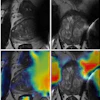
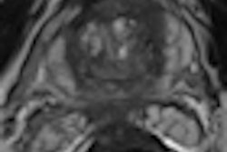
While 70% of prostate cancer originates in the prostate's peripheral zone, 30% stem from the central gland. Finding these cancers from the central gland on MRI is a challenging task, but Dutch researchers believe their computer-aided diagnosis (CADx) tool can help.
A team from Radboud University Nijmegen Medical Centre's Diagnostic Image Analysis Group found their CADx system yielded similar performance to that of radiologists reported in the literature. Geert Litjens traveled to Chicago to present the team's findings at the 2012 RSNA meeting, and our coverage of his presentation can be found here.
Be sure also to check back in your Advanced Visualization Digital Community in the coming weeks for additional coverage of European advanced visualization research presented at the RSNA meeting.
Mammography computer-aided detection (CAD) research figures prominently in other articles featured in this issue. A Spanish team recently found that CAD can offer high sensitivity for finding very small invasive breast cancers on full-field digital mammography. As has often been the case with CAD, the software did suffer from low specificity in their study, however. Get all the details here.
Other Dutch research determined that radiologists can be more accurate at detecting malignant breast masses if CAD marks are obscured until they are needed, rather than shown during reading. The group found that an interactive CAD system that provides results on-demand for queried regions led to a higher sensitivity for radiologists than traditional CAD prompts.
Speaking of CAD, our columnist Dr. Peter Rinck warns how getting used to CAD can make radiologists lazy in a recent edition of the Maverinck.
Happy holidays from all of us at AuntMinnieEurope.com!







![A normal mammogram confirmed by three-year radiologic follow-up illustrates reader-marked regions of interest (ROIs) during (A) unaided (round 1) and (B) artificial intelligence (AI)–assisted (round 2) reading. Each colored dot represents an ROI for recall by a human reader. Readers could mark more than one ROI per case, represented by multiple dots of the same color. During AI-assisted reading, the AI system displayed three visible prompts: two with suspicion of malignancy scores of 35% (left mediolateral oblique [L MLO] and craniocaudal [L CC]) and one with a suspicion of malignancy score of 10% (right craniocaudal [R CC]), shown as polygonal overlays. Without AI, six of 10 readers (60%) marked a false-positive ROI. With AI assistance, this fell to two of 10 (20%). R MLO = right mediolateral oblique.](https://img.auntminnieeurope.com/mindful/smg/workspaces/default/uploads/2026/07/2026-07-14-radiology-mammogram-ai-auto-bias.H0bYO8QlWs.jpg?auto=format%2Ccompress&dpr=2&fit=crop&h=167&q=70&w=250)


