Dear Women's Imaging Insider,
It's widely known that device-related factors, including acquisition and system parameters, have an impact on image quality. However, relatively little is known about the specific influence of different mammography systems on breast density evaluations.
To address this shortfall, Belgian researchers have conducted a timely study comparing the performance of scanners from three leading vendors, and they've just published their findings in European Radiology. Find out more in today's top story.
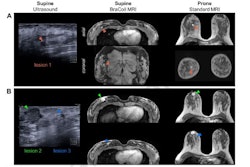
Meanwhile, an Austrian study has found that supine breast MRI with a lightweight, wearable radiofrequency coil vest can help to improve lesion localization in radiotherapy planning, surgery, targeted ultrasound, and ultrasound-guided biopsy, potentially reducing the need for MR-guided biopsies.
In another important recent paper, the authors found that the presence of an oval-shaped mass and posterior shadowing are significant imaging markers for assessing benign breast lesions categorized as BI-RADS 4c or 5.
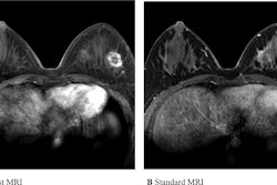
Our editorial team has also been impressed by the work of a Korean group that found that an ultrafast MRI radiomics model matched the performance of standard MRI in classifying hormone receptors, HER2 status, and molecular subtypes.
Thankfully, pathologies of the vulva are rare -- but because of their rareness, many radiologists struggle to diagnose some conditions correctly. This has prompted a research team from Vienna and Rome to produce an authoritative and comprehensive article on MRI of the vulva.
Philip Ward
Editor in Chief
AuntMinnieEurope.com




![A normal mammogram confirmed by three-year radiologic follow-up illustrates reader-marked regions of interest (ROIs) during (A) unaided (round 1) and (B) artificial intelligence (AI)–assisted (round 2) reading. Each colored dot represents an ROI for recall by a human reader. Readers could mark more than one ROI per case, represented by multiple dots of the same color. During AI-assisted reading, the AI system displayed three visible prompts: two with suspicion of malignancy scores of 35% (left mediolateral oblique [L MLO] and craniocaudal [L CC]) and one with a suspicion of malignancy score of 10% (right craniocaudal [R CC]), shown as polygonal overlays. Without AI, six of 10 readers (60%) marked a false-positive ROI. With AI assistance, this fell to two of 10 (20%). R MLO = right mediolateral oblique.](https://img.auntminnieeurope.com/mindful/smg/workspaces/default/uploads/2026/07/2026-07-14-radiology-mammogram-ai-auto-bias.H0bYO8QlWs.jpg?auto=format%2Ccompress&fit=crop&h=100&q=70&w=100)


![A normal mammogram confirmed by three-year radiologic follow-up illustrates reader-marked regions of interest (ROIs) during (A) unaided (round 1) and (B) artificial intelligence (AI)–assisted (round 2) reading. Each colored dot represents an ROI for recall by a human reader. Readers could mark more than one ROI per case, represented by multiple dots of the same color. During AI-assisted reading, the AI system displayed three visible prompts: two with suspicion of malignancy scores of 35% (left mediolateral oblique [L MLO] and craniocaudal [L CC]) and one with a suspicion of malignancy score of 10% (right craniocaudal [R CC]), shown as polygonal overlays. Without AI, six of 10 readers (60%) marked a false-positive ROI. With AI assistance, this fell to two of 10 (20%). R MLO = right mediolateral oblique.](https://img.auntminnieeurope.com/mindful/smg/workspaces/default/uploads/2026/07/2026-07-14-radiology-mammogram-ai-auto-bias.H0bYO8QlWs.jpg?auto=format%2Ccompress&fit=crop&h=112&q=70&w=112)