Dear Women's Imaging Insider,
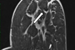
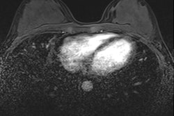
What's the impact of metallic markers on the accuracy and interpretation of breast MRI scans?
Swiss researchers have investigated this important -- but rarely studied -- question. Although their sample size was relatively small and they only studied two different types of markers, their results and analysis deserve a close look.
In our second article posted today, a team from Paris reported that MRI with an ultrafast dynamic contrast-enhanced protocol can help to predict complete pathologic response to breast cancer treatment. The group found that by measuring the slope between the time of contrast inflow onset and the time of peak intensity in breast tumors, known as the wash-in slope, clinicians can predict how tumors respond to neoadjuvant therapy with high sensitivity.
Another recent story on breast MRI asked whether a clinical decision algorithm can outweigh reader experience. A highly respected research group from Vienna has investigated this topic, and the team published its results in European Radiology.
Breast density continues to be a challenge for radiologists, but research from the U.K. and the Netherlands sheds new light on how imaging professionals can refine cancer risk estimates for women with dense breast tissue. Find out more in this news report from ECR 2022 in Vienna.
In other news, Italian investigators have concluded that mammograms performed with women compressing their own breasts have comparable image quality and significantly stronger breast compression.
In this newsletter, we've singled out a small selection of the diverse range of articles posted in the Women's Imaging Community over the past month or so. Please scroll through the full list below, and feel free to contact me if you have ideas for future coverage.



![A normal mammogram confirmed by three-year radiologic follow-up illustrates reader-marked regions of interest (ROIs) during (A) unaided (round 1) and (B) artificial intelligence (AI)–assisted (round 2) reading. Each colored dot represents an ROI for recall by a human reader. Readers could mark more than one ROI per case, represented by multiple dots of the same color. During AI-assisted reading, the AI system displayed three visible prompts: two with suspicion of malignancy scores of 35% (left mediolateral oblique [L MLO] and craniocaudal [L CC]) and one with a suspicion of malignancy score of 10% (right craniocaudal [R CC]), shown as polygonal overlays. Without AI, six of 10 readers (60%) marked a false-positive ROI. With AI assistance, this fell to two of 10 (20%). R MLO = right mediolateral oblique.](https://img.auntminnieeurope.com/mindful/smg/workspaces/default/uploads/2026/07/2026-07-14-radiology-mammogram-ai-auto-bias.H0bYO8QlWs.jpg?auto=format%2Ccompress&fit=crop&h=100&q=70&w=100)


![A normal mammogram confirmed by three-year radiologic follow-up illustrates reader-marked regions of interest (ROIs) during (A) unaided (round 1) and (B) artificial intelligence (AI)–assisted (round 2) reading. Each colored dot represents an ROI for recall by a human reader. Readers could mark more than one ROI per case, represented by multiple dots of the same color. During AI-assisted reading, the AI system displayed three visible prompts: two with suspicion of malignancy scores of 35% (left mediolateral oblique [L MLO] and craniocaudal [L CC]) and one with a suspicion of malignancy score of 10% (right craniocaudal [R CC]), shown as polygonal overlays. Without AI, six of 10 readers (60%) marked a false-positive ROI. With AI assistance, this fell to two of 10 (20%). R MLO = right mediolateral oblique.](https://img.auntminnieeurope.com/mindful/smg/workspaces/default/uploads/2026/07/2026-07-14-radiology-mammogram-ai-auto-bias.H0bYO8QlWs.jpg?auto=format%2Ccompress&fit=crop&h=112&q=70&w=112)