Dear Women's Imaging Insider,
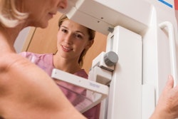
Mammography recall is a vitally important area of women's imaging, but there's been relatively little investigation into optimal recall rates. That's why a new analysis of more than 11 million breast screening examinations from the English National Health Service (NHS) program is bound to generate considerable interest, particularly because the study was conducted by an experienced and respected group of researchers.
Significantly, the investigators' methods may help countries with mammography screening programs to determine their optimal recall rates, as well as help the English scheme set its own recall rate target. Make sure you check out their findings.
Should regular screening of women identified with a moderate or high risk of breast cancer be extended from ages 40 to 49 to also include ages 35 to 39? This question has come under intense scrutiny over recent weeks.
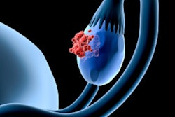
Meanwhile, swift progress continues to be made in the use of artificial intelligence (AI) in women's imaging. An international group of investigators has developed an AI algorithm capable of predicting the survival rate of patients with ovarian cancer by analyzing their contrast-enhanced CT scans.
Much more work needs to be done to avoid the risk of cross-contamination in endovaginal ultrasound examinations, Israeli authors write. The Israeli Ministry of Health has issued four practical suggestions to help improve the situation.
If you have any ideas or advice for topics you'd like to see covered in the Women's Imaging Community, please feel free to get in touch.



![A normal mammogram confirmed by three-year radiologic follow-up illustrates reader-marked regions of interest (ROIs) during (A) unaided (round 1) and (B) artificial intelligence (AI)–assisted (round 2) reading. Each colored dot represents an ROI for recall by a human reader. Readers could mark more than one ROI per case, represented by multiple dots of the same color. During AI-assisted reading, the AI system displayed three visible prompts: two with suspicion of malignancy scores of 35% (left mediolateral oblique [L MLO] and craniocaudal [L CC]) and one with a suspicion of malignancy score of 10% (right craniocaudal [R CC]), shown as polygonal overlays. Without AI, six of 10 readers (60%) marked a false-positive ROI. With AI assistance, this fell to two of 10 (20%). R MLO = right mediolateral oblique.](https://img.auntminnieeurope.com/mindful/smg/workspaces/default/uploads/2026/07/2026-07-14-radiology-mammogram-ai-auto-bias.H0bYO8QlWs.jpg?auto=format%2Ccompress&fit=crop&h=100&q=70&w=100)


![A normal mammogram confirmed by three-year radiologic follow-up illustrates reader-marked regions of interest (ROIs) during (A) unaided (round 1) and (B) artificial intelligence (AI)–assisted (round 2) reading. Each colored dot represents an ROI for recall by a human reader. Readers could mark more than one ROI per case, represented by multiple dots of the same color. During AI-assisted reading, the AI system displayed three visible prompts: two with suspicion of malignancy scores of 35% (left mediolateral oblique [L MLO] and craniocaudal [L CC]) and one with a suspicion of malignancy score of 10% (right craniocaudal [R CC]), shown as polygonal overlays. Without AI, six of 10 readers (60%) marked a false-positive ROI. With AI assistance, this fell to two of 10 (20%). R MLO = right mediolateral oblique.](https://img.auntminnieeurope.com/mindful/smg/workspaces/default/uploads/2026/07/2026-07-14-radiology-mammogram-ai-auto-bias.H0bYO8QlWs.jpg?auto=format%2Ccompress&fit=crop&h=112&q=70&w=112)