Dr. Fabian Kiessling is one of the very few European radiologists who have truly embraced the brave new world of molecular medicine. For the past four years, he has been chairman of experimental molecular imaging at RWTH-Aachen University in Germany, and he is joint head of the educational committee for the World Molecular Imaging Congress (WMIC), which starts today in Dublin.
He was previously junior group leader for molecular imaging at the German Cancer Research Center (DKFZ) in Heidelberg, where he undertook his radiology training. In 2008, Kiessling founded InvivoContrast with Dr. Matthias Braeutigam, former head of imaging diagnostic research at Bayer Schering Pharma, Berlin. The company aims to support the imaging research community with easily accessible and reliable contrast agents for preclinical research.
To coincide with the start of the WMIC, we interviewed Kiessling about the current status and future development of molecular imaging (MI). Further news stories from the congress will follow over the next few days.
 There is a definite need for more money for production of new tracers for clinical application and for early clinical trials, according to Dr. Fabian Kiessling.
AuntMinnieEurope.com: Of the various definitions of MI, which do you like most?
Kiessling: I prefer the original one from Dr. Douglas Wagenaar et al since this one is most precise. They defined MI as "the in vivo characterization and measurement of biologic processes at the cellular and molecular levels." They also noted that this "growing research discipline is aimed at developing and testing novel tools, reagents, and methods to image specific molecular pathways in vivo -- particularly those that are key targets in disease processes" (Academic Radiology, 2001 May, Vol. 8:55, pp. 409-420).
Personally, though, I don't really like the term "molecular imaging" because we're doing so much than just that.
Which areas of MI offer the greatest future clinical potential?
The use of targeted probes for therapy personalization since the motivation of the industry is high to implement this. Also, I think PET/MRI will eventually become a cost-effective technique in routine clinical use because you can save space and time, and attract more patients.
Are general radiologists sufficiently interested in MI?
There is significant interest. However, there is still not enough willingness to change the mind-set and to strongly consider pathophysiology and molecular processes in radiologists' diagnostic conduct. Radiologists have to basically understand how each molecular probe describes a biological pathway and how the pathway is linked to the disease.
Overall, do you consider Europe lagging behind the U.S. in MI?
Considering the number of scientists working on MI and the availability of funding, I think Europe is not too much behind the U.S. The U.S. might be stronger in bringing new ideas into clinics and making products out of it. However, there is never enough funding for research, and particular programs dedicated to MI would be helpful. There is a definite need for more money for production of new tracers for clinical application and for early clinical trials.
 |
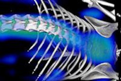
| Fusion of ultrashort-echo-time (UTE) MR image (gray; TE = 0.09 sec) and smoothed PET image (orange). PET data attenuation-corrected using UTE triple-echo MR data. From left to right, transaxial, coronal, sagittal cross-sections. Method from Berker et al, Journal of Nuclear Medicine, May 2012, Vol. 53:5, pp. 796-804. |
How do you organize your own department's research program?
We have five different groups devoted to "physics and engineering of imaging modalities, particularly on PET/MRI," "MI probe development," "tumor angiogenesis and microenvironment," "nanomedicines and theranostics," and "clinical translation and late preclinical studies." There is now a total staff of 43, comprising three radiologists, a pathologist, a cardiologist, 10 physicists and engineers, 10 biologists (including doctoral candidates), five chemists, two pharmacologists, and several informatics specialists and technicians. We're a really multinational and multidisciplinary team!
Only few radiologists are really interested in joining a department like ours, but then again we don't need many of them. We mainly need excellent clinicians. In addition, the research about molecular imaging is complicated, and people need to learn much about physics, chemistry, and biology. Therefore, a long-term research commitment is usually required.
We're looking for very curious people that clearly aim at an academic career. Basic research has certain risks, however. We tend not to give clinical colleagues high-risk projects because it would be a disaster for them if they spent a half year on a project and it didn't work out.
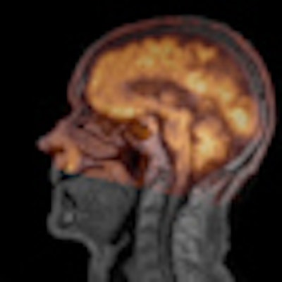
 Silicon photomultiplier based preclinical PET/RF insert for simultaneous PET and MR imaging in a human 3-tesla MRI at University Clinic in Aachen. Presented by V. Schulz et al, IEEE Nuclear Science Symposium and Medical Imaging Conference, 2011, rec. J2-5.
Could the European Society of Molecular Imaging (ESMI) and European Association of Nuclear Medicine (EANM) eventually merge?
No, I don't think the two groups will ever merge. ESMI is much more multidisciplinary (modalities: MRI, ultrasound, optical imaging, PET, SPECT, magnetic particle imaging; fields: nanotechnology, small probes, biochemistry, etc.) and is more focused on basic research (e.g., also on developmental biological processes). EANM focuses on nuclear medicine and its clinical applications, and most of its members are nuclear medicine doctors and radiochemists. ESMI's members are scientists coming from all fields. Of course there is some overlap between them, however.
Are you optimistic about the long-term future of MI?
Of course. If not, I would switch to another field.