German developers of dark-field chest x-ray appear to have overcome a technical limitation of the technology – namely, adjusting for photon scattering caused by interferometers used in the experimental system.
This scattering is picked up by the system’s detector (so-called “detector cross talk”) and leads to unwanted artifacts on patient chest x-rays, noted study lead and doctoral candidate Theresa Urban, of the Technical University of Munich, and colleagues. Ultimately, the group described a method to correct the phenomenon to produce better images.
“With the corrections presented here … the obtained dark-field signal is due to the microstructure of the tissue, and differences between the dark-field signal of different patients can be attributed to their lung condition rather than scatter or crosstalk artifacts,” the group wrote. The article was published on 7 March in IEEE Transactions on Medical Imaging.
Initial results from currently ongoing clinical studies suggest that dark-field chest x-ray can be useful for detecting and quantification of pulmonary emphysema, and for assessing COVID-19 pneumonia, the authors wrote.
The technology uses three interferometers in the path of an x-ray beam from a conventional system to focus photons on the lung’s alveoli, or microscopic structures located at the end of the respiratory tree. Yet photon scattering and detector cross talk cause artifacts in the dark-field x-ray image, which impairs image appearance and prevents a quantitative analysis of the dark-field signal, the team wrote.
To overcome this limitation, the researchers developed a deconvolution-based correction method for the induced artifacts.
In brief, Urban and colleagues developed a method to calculate what are known as scattering “kernels,” or mathematical descriptions of how the photons scatter when they interact with molecules. These kernels were then used as input for an algorithm that corrected for them in the output – in other words, ultimately sharpening the resolution of the x-ray, they explained.
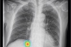
With the physics elucidated, the researchers then validated the correction method with a water phantom and finally obtained successful correction on dark-field images of a human thorax.
“This ensures an unobstructed qualitative evaluation, and enables a quantitative evaluation of dark-field radiographs,” Urban et al wrote.
Dark-field chest x-ray has just recently been translated to the clinical stage and its diagnostic value is currently being investigated with a clinical prototype, the authors wrote. They noted that the overall scatter that reaches the detector depends on the beam spectrum and the distance from the x-ray beam to the target, and the influence of changes in these parameters will require further investigations, they wrote.
“However, the general concepts behind the corrections presented here are applicable for all grating-based radiography systems, including even the recently presented clinical dark-field CT system,” the researchers concluded.
Access to the full article is available here.