Using a simulator app in radiation protection training can boost understanding of x-ray scatter in different imaging scenarios, researchers from Finland have found.
Conventional projection radiography remains the most common x-ray-based imaging modality, but interventional radiology examinations have increased considerably over the last 15 years, Satu Ylimaula, a doctoral candidate at the Research Unit of Health Sciences and Technology at the University of Oulu and medical physics resident at the Department of Diagnostic Radiology at Oulu University Hospital, and colleagues explained.
To minimize occupational exposure to scattered radiation, healthcare professionals in the European Union receive continuous radiation protection training based on Directive 2013/59/Euratom. Various methods can be used in the training to visualize scattered radiation, they noted.
 The research process. All figures courtesy of Satu Ylimaula et al and ESR EPOS database.
In their study presented as a poster at EuroSafe 2025, the researchers measured x-ray scatter using several imaging protocols. Based on the measurements, they designed and created x-ray scatter maps and, subsequently, a mobile application for radiation protection training purposes. A user experience study of the app is ongoing.
The group used an ATOM dosimetry verification phantom 701/C from Sun Nuclear as a scattering object, and an active dosimetry system (Raysafe i2, Unfors Raysafe AB) was used to measure the dose rate of the scattered x-ray radiation at various locations around the scattering object.
Matlab R2022b -- a programming and numeric computing platform from Mathworks that can analyze data, develop algorithms, and create models -- was used to generate scattered radiation maps. The Oulu team used Blender 3.4 software from Blender Foundation to transfer maps into 3D models, while Unity 2021.3.12f from Unity Technologies was used to create an x-ray scatter simulator mobile app that presents these 3D maps in augmented reality.
 X-ray scatter app in use.
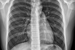
The app includes scatter maps for these x-ray imaging protocols: posterior-anterior (PA) chest x-rays (standing), anterior-posterior (AP) mobile chest x-rays (supine), and urological procedures utilizing fluoroscopy.
The researchers invited radiography students and healthcare radiation workers to use the app and participate in a user experience study. The user experience study is ongoing, with radiography students comprising three-quarters of the total participants (n = 59) so far.
“Based on our initial observations, the user experience of the simulator varies,” they noted. “In addition to the actual app design and personal preferences, possible reasons for the variation could include differences in software, camera, and the physical operating environment of the device the app is used on.”
The majority of participants reported that the app enhanced their understanding of x-ray scatter behavior in different imaging protocols, and they felt it was a valuable addition to traditional radiation protection training. Furthermore, completing small tasks by users, such as calculating potential exposure levels in different imaging protocols and locations within the imaging room, can maximize the app’s potential and further improve understanding of exposure differences in various imaging situations, the authors wrote.
 QR codes to the app’s download pages in the Play Store and the App Store.
You can read the full e-poster on the EPOS database of the ESR. The work was presented at Eurosafe 2025. The co-authors were Vili Tuppurainen, Nina Hänninen, Karoliina Paalimäki-Paakki, Lasse Räsänen, Miika Tapio Nieminen, and Matti Hanni.
For further reading, go to this article by the same authors: X-ray scatter in projection radiography, Radiation Protection Dosimetry, vol. 200, no. 2, pp.120-129, February 2024.