When breath-holding is a problem in patients with nonalcoholic fatty liver disease, MRI practitioners need to think more deeply about modifying their technique, according to research published on 21 March in European Radiology.
3D chemical shift-encoded (CSE) MRI enables accurate and precise quantification of proton density fat fraction (PDFF) and R2* -- two important biomarkers of hepatic fat and iron deposition -- but 3D CSE-MRI requires reliable breath-holding, noted lead authors Dr. Jitka Starekova and Ruiyang Zhao from the University of Wisconsin, Madison, U.S.
"Breath-holding is difficult for some children and adults, including those with respiratory disease, cognitive impairment, and/or hearing loss. As a result, motion artifacts, which can lead to aliasing of signal from adipose tissue into the liver, introduce bias and variability in PDFF and R2* measurements," they explained.
Free-breathing 2D CSE-MRI with sequential radiofrequency excitation is a motion-robust alternative, but it suffers from low signal-to-noise ratio (SNR). To overcome this limitation, the researchers decided to look at the combination of flip angle-modulated (FAM) 2D CSE imaging with a nonlocal means (NLM) motion-corrected averaging technique.
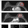
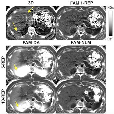
 The free-breathing multi-repetition 2D chemical shift-encoded (CSE) flip angle modulation (FAM) nonlocal means (NLM) R2* maps showed significant improvement in signal-to-noise ratio (SNR) and reduction of artifacts. Shown here are R2* maps of an 18-year-old male patient with an R2* of 41 s-1. Motion artifacts are depicted by dashed arrows. Of note are signal inhomogeneities (arrows) in the right lobe with multi-repetition FAM using direct averaging (DA), which lead to bias. Note the improved image quality and higher SNR with increasing number of FAM repetitions (REP = number of repetitions). Representative scores for SNR, artifacts, and overall image quality are 3,2,2 (3D); 3,5,4 (FAM 1- REP); 5,2,2 (FAM-DA 5-REP); 4,5,4 (FAM-NLM 5-REP); 5,2,3 (FAM-DA 10-REP); and 5,5,5 (FAM-NLM 10-REP). Image courtesy of Dr. Jitka Starekova, Ruiyang Zhao et al.; University of Wisconsin; and European Radiology.
In a prospective study, the Wisconsin group scanned 35 healthy subjects (27 children, 8 adults) using a 3T MRI system (Discovery MR750, GE Healthcare) and a 32-channel phased-array torso coil (Neocoil, GE Healthcare). The breath-hold 3D acquisition time was approximately 20 seconds, while that for free-breathing FAM was approximately 26 seconds per repetition (acquisition time for the 10-repetition FAM acquisitions was 260 seconds).
The team acquired multi-echo 3D CSE and 2D CSE FAM images during breath-hold and free-breathing, respectively, to obtain PDFF and R2* maps of the liver. Multi-repetition FAM was postprocessed with direct averaging (DA) and NLM-based averaging and compared with 3D CSE using Bland-Altmann and regression analysis. Image qualities of PDFF and R2* maps were reviewed by two radiologists using a Likert-like scale (score 1-5, 5 = best).
Compared with 3D CSE, multirepetition FAM-NLM showed excellent agreement (regression slope = 1.0, R2 = 0.996) for PDFF and good agreement (regression slope 1.08-1.15, R2 ≥ 0.899) for R2*. Also, multirepetition FAM-NLM PDFF and R2* maps had fewer artifacts (score 3.8 vs. 3.2, p < 0.0001 for PDFF; score 3.2 vs. 2.6, p < 0.001 for R2*) and better overall image quality (score 4.0 vs. 3.5, p < 0.0001 for PDFF; score 3.4 vs. 2.7, p < 0.0001 for R2*).
Impact on clinical practice
Overall, free-breathing FAM-NLM provides superior image quality of the liver compared with the conventional breathhold 3D CSE-MRI, while minimizing bias for PDFF and R2* quantification, the authors stated.
The results were generally in line with expectations, corresponding author Dr. Diego Hernando told AuntMinnieEurope.com. For MRI-based liver fat and R2* quantification, the combination of optimized “flip-angle modulated” acquisitions during free-breathing -- along with multiple repetitions that are combined using motion-corrected averaging -- leads to synergistic gains in SNR, he said.
The team's main hope is the study will provide a significant boost for free-breathing quantitative MRI of diffuse liver disease.
"We expect these developments to have an impact on the imaging of patients who are unable to complete a breath-hold," Hernando commented. "More broadly, the ability to perform fully free-breathing quantitative MRI of the abdomen may lead to improvements in workflow and efficiency for clinical imaging."
Future plans and ISMRM 2022
The plan now is to perform further studies on the proposed methods, including optimization of the acquisition and reconstruction methods. In addition, the researchers will validate the proposed high-SNR, free-breathing methods in specific patients, such as those with iron overload.
They will also present multiple recent developments at the annual congress of the International Society of Magnetic Resonance in Medicine (ISMRM 2022), to be held in London in May. For example, a study led by Ruiqi Geng (AI-based automated liver image prescription: evaluation across patients and pathologies and prospective implementation and validation) has demonstrated the performance of an automated method for image prescription in liver imaging.
"This liver image prescription method is highly complementary to the free-breathing fat and R2* mapping method reported in the European Radiology manuscript, as the combination of these techniques may open the door to fully-automated 'single button-push' MRI of the liver for the evaluation of diffuse liver disease," Hernando said.