A fast-scanning cardiac MRI protocol first developed and tested in the U.K. has been successfully used in Peru, according to research published on 29 August in the Journal of the American Heart Association. Researchers believe the study proves the technique is well-suited for providing cardiac MRI in developing countries.
Cardiac MRI is an emerging modality that has great potential to change the care of patients with heart disease, but there are multiple techniques available to measure parameters such as function, scarring, and perfusion. These techniques make the modality slow, with scans taking as long as 45 minutes, and too expensive and complex for most developing countries.
For example, in Peru, there are only two MRI scanners that perform cardiac MRI; they provide a total of six scans per week at a cost of $400 (345 euros) to $800 (689 euros) per scan. What if a lower-cost, faster protocol were developed?
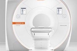
To that end, an international group of researchers developed a cardiac MR protocol for a 1.5-tesla scanner to acquire data on cardiac volume, function, and scarring based on late gadolinium enhancement. The rapid protocol can be performed on existing MRI scanners, so no significant hardware upgrades are required.
To find out how it worked in a developing country, the approach was evaluated on 98 cardiac patients (average age, 52) in Peru in the Impact of Noninvasive CMR Assessment (INCA) study. The scanning technique showed hypertrophic cardiomyopathy in 26% of patients, dilated cardiomyopathy in 22%, and ischemic cardiomyopathy in 15%.
In addition, the scans revealed 12 other pathologies, including tumors, congenital heart disease, iron overload, amyloid plaques, inflamed vessels, clots, and valve disease. The technique also led to a new, unsuspected diagnosis in 19% of the patients, while treatment plans were changed to a more appropriate course in 37%.
The exams were completed in 18 minutes, compared with 45 minutes for conventional scans, and cost approximately $150 per patient.
"Our cardiac MR strategy was three to five times cheaper than current cardiac MR exams in Peru," said lead author Dr. James Moon, a professor at Barts Heart Centre at St. Bartholomew's Hospital in London. "It also can be delivered two to three times faster and is easier than conventional cardiac MR."
More information on the rapid cardiac MR technique is available by clicking here.


![Examples of ultrasound findings and techniques. (A) Images in a 39-year-old male patient with a mass in the left thigh. The mass is heterogeneous on the B-mode US image (compared with the patient in D) and showed increased microvascularity (superb microvascular imaging [SMI]) and shear-wave elastography (SWE) values. Undifferentiated pleomorphic sarcoma was diagnosed at biopsy (with pleomorphic rhabdomyosarcoma in surgical specimen). (B) Images in an 18-year-old male patient with a mass in the left leg. The mass is hypoechoic on the B-mode image, with no other findings suggestive of malignancy. The lesion is in contact with the cortex of the tibia, which is slightly irregular. CT revealed a doubtful anteromedial tibial erosion. The microvascular study demonstrated high vascularization, suggestive of malignancy. Periosteal Ewing sarcoma was diagnosed with both histologic and immunohistochemical confirmation. (C) Images in a 69-year-old female patient with a lump growing on the outside of the left leg. Multiple SWE examinations were performed (please note the high values obtained in the measurements, whereas the color map highlights the stiffness relative to adjacent tissues). SMI showed areas of increased vascularization to target for sampling. Undifferentiated spindle cell sarcoma was diagnosed at biopsy, with residual leiomyosarcoma in the surgical specimen after neoadjuvant therapy. (D) Images in a 56-year-old female patient with a mass in the right thigh. The mass is heterogeneous at both B-mode ultrasound (similar to patient A) and MRI (coronal T2-weighted spectral attenuated inversion recovery [SPAIR]; T1-weighted pre-contrast and postcontrast imaging), which even shows uptake after the administration of paramagnetic contrast material, which is traditionally suggestive of malignancy. Low values at SMI and elastography are suggestive of benignity. Spindle cell lipoma was diagnosed at biopsy, with atypical spindle cell lipomatous tumor in the surgical specimen.](https://img.auntminnieeurope.com/mindful/smg/workspaces/default/uploads/2026/08/images-radiol250278fig2.APCFLSvX6p.jpg?auto=format%2Ccompress&fit=crop&h=100&q=70&w=100)


![Examples of ultrasound findings and techniques. (A) Images in a 39-year-old male patient with a mass in the left thigh. The mass is heterogeneous on the B-mode US image (compared with the patient in D) and showed increased microvascularity (superb microvascular imaging [SMI]) and shear-wave elastography (SWE) values. Undifferentiated pleomorphic sarcoma was diagnosed at biopsy (with pleomorphic rhabdomyosarcoma in surgical specimen). (B) Images in an 18-year-old male patient with a mass in the left leg. The mass is hypoechoic on the B-mode image, with no other findings suggestive of malignancy. The lesion is in contact with the cortex of the tibia, which is slightly irregular. CT revealed a doubtful anteromedial tibial erosion. The microvascular study demonstrated high vascularization, suggestive of malignancy. Periosteal Ewing sarcoma was diagnosed with both histologic and immunohistochemical confirmation. (C) Images in a 69-year-old female patient with a lump growing on the outside of the left leg. Multiple SWE examinations were performed (please note the high values obtained in the measurements, whereas the color map highlights the stiffness relative to adjacent tissues). SMI showed areas of increased vascularization to target for sampling. Undifferentiated spindle cell sarcoma was diagnosed at biopsy, with residual leiomyosarcoma in the surgical specimen after neoadjuvant therapy. (D) Images in a 56-year-old female patient with a mass in the right thigh. The mass is heterogeneous at both B-mode ultrasound (similar to patient A) and MRI (coronal T2-weighted spectral attenuated inversion recovery [SPAIR]; T1-weighted pre-contrast and postcontrast imaging), which even shows uptake after the administration of paramagnetic contrast material, which is traditionally suggestive of malignancy. Low values at SMI and elastography are suggestive of benignity. Spindle cell lipoma was diagnosed at biopsy, with atypical spindle cell lipomatous tumor in the surgical specimen.](https://img.auntminnieeurope.com/mindful/smg/workspaces/default/uploads/2026/08/images-radiol250278fig2.APCFLSvX6p.jpg?auto=format%2Ccompress&fit=crop&h=112&q=70&w=112)