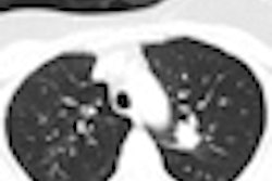
Advanced radiotherapy techniques are attributed to improvements in survival of elderly lung cancer patients in the Netherlands, according to a new study published online on 2 May in Annals of Oncology.
Researchers at the VU University Medical Center in Amsterdam conducted a population-based study evaluating time trends in treatment patterns and survival of 4,605 patients over the age of 75 diagnosed with early-stage lung cancer. Comparisons were made between three consecutive time periods: from 2001-2003, from 2004-2006, and from 2007-2009.
Lead author Dr. Niels Haasbeek from the department of radiation oncology reported that between 2007 and 2009, when stereotatic ablative radiotherapy was available in all of the country's cancer treatment centers, and the first time period, when this treatment was not available, patients survived a median of nearly 10 additional months.
During the first time period, the median survival rate was 16.8 months following treatment. With the availability of stereotatic ablative radiotherapy (SABR) treatment, patients in the most recent time period lived 26.1 months.
"The greater use of advanced radiotherapy techniques has led to large improvements in survival for Dutch lung cancer patients over the age of 75, many of whom are too frail to undergo surgery," Haasbeek noted. "Those patients in this age group who are fit enough for surgery should also be informed about the curative option of SABR as an alternative to surgery."
SABR -- sometimes called stereotactic body radiotherapy, or SBRT -- refers to a method of treating the tumor with a targeted high-energy radiation beam to damage tumor cell DNA and kill the cancer cell. It was first used in the Netherlands at the VUMC in 2003. The researchers used the Dutch National Cancer Registry to examine survival data for three defined periods: the three-year period before it was introduced, the three-year period while it was becoming available at other Dutch centers, and the three years when it was available nationwide, according to Dr. Suresh Senan, radiation oncologist at VUMC, who led the study based on nationwide data.
"We noted a marked improvement in survival among the nearly 5,000 lung cancer patients aged over 75 who were treated over these nine years," Senan concluded.


![Examples of ultrasound findings and techniques. (A) Images in a 39-year-old male patient with a mass in the left thigh. The mass is heterogeneous on the B-mode US image (compared with the patient in D) and showed increased microvascularity (superb microvascular imaging [SMI]) and shear-wave elastography (SWE) values. Undifferentiated pleomorphic sarcoma was diagnosed at biopsy (with pleomorphic rhabdomyosarcoma in surgical specimen). (B) Images in an 18-year-old male patient with a mass in the left leg. The mass is hypoechoic on the B-mode image, with no other findings suggestive of malignancy. The lesion is in contact with the cortex of the tibia, which is slightly irregular. CT revealed a doubtful anteromedial tibial erosion. The microvascular study demonstrated high vascularization, suggestive of malignancy. Periosteal Ewing sarcoma was diagnosed with both histologic and immunohistochemical confirmation. (C) Images in a 69-year-old female patient with a lump growing on the outside of the left leg. Multiple SWE examinations were performed (please note the high values obtained in the measurements, whereas the color map highlights the stiffness relative to adjacent tissues). SMI showed areas of increased vascularization to target for sampling. Undifferentiated spindle cell sarcoma was diagnosed at biopsy, with residual leiomyosarcoma in the surgical specimen after neoadjuvant therapy. (D) Images in a 56-year-old female patient with a mass in the right thigh. The mass is heterogeneous at both B-mode ultrasound (similar to patient A) and MRI (coronal T2-weighted spectral attenuated inversion recovery [SPAIR]; T1-weighted pre-contrast and postcontrast imaging), which even shows uptake after the administration of paramagnetic contrast material, which is traditionally suggestive of malignancy. Low values at SMI and elastography are suggestive of benignity. Spindle cell lipoma was diagnosed at biopsy, with atypical spindle cell lipomatous tumor in the surgical specimen.](https://img.auntminnieeurope.com/mindful/smg/workspaces/default/uploads/2026/08/images-radiol250278fig2.APCFLSvX6p.jpg?auto=format%2Ccompress&fit=crop&h=100&q=70&w=100)


![Examples of ultrasound findings and techniques. (A) Images in a 39-year-old male patient with a mass in the left thigh. The mass is heterogeneous on the B-mode US image (compared with the patient in D) and showed increased microvascularity (superb microvascular imaging [SMI]) and shear-wave elastography (SWE) values. Undifferentiated pleomorphic sarcoma was diagnosed at biopsy (with pleomorphic rhabdomyosarcoma in surgical specimen). (B) Images in an 18-year-old male patient with a mass in the left leg. The mass is hypoechoic on the B-mode image, with no other findings suggestive of malignancy. The lesion is in contact with the cortex of the tibia, which is slightly irregular. CT revealed a doubtful anteromedial tibial erosion. The microvascular study demonstrated high vascularization, suggestive of malignancy. Periosteal Ewing sarcoma was diagnosed with both histologic and immunohistochemical confirmation. (C) Images in a 69-year-old female patient with a lump growing on the outside of the left leg. Multiple SWE examinations were performed (please note the high values obtained in the measurements, whereas the color map highlights the stiffness relative to adjacent tissues). SMI showed areas of increased vascularization to target for sampling. Undifferentiated spindle cell sarcoma was diagnosed at biopsy, with residual leiomyosarcoma in the surgical specimen after neoadjuvant therapy. (D) Images in a 56-year-old female patient with a mass in the right thigh. The mass is heterogeneous at both B-mode ultrasound (similar to patient A) and MRI (coronal T2-weighted spectral attenuated inversion recovery [SPAIR]; T1-weighted pre-contrast and postcontrast imaging), which even shows uptake after the administration of paramagnetic contrast material, which is traditionally suggestive of malignancy. Low values at SMI and elastography are suggestive of benignity. Spindle cell lipoma was diagnosed at biopsy, with atypical spindle cell lipomatous tumor in the surgical specimen.](https://img.auntminnieeurope.com/mindful/smg/workspaces/default/uploads/2026/08/images-radiol250278fig2.APCFLSvX6p.jpg?auto=format%2Ccompress&fit=crop&h=112&q=70&w=112)