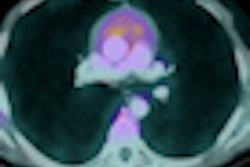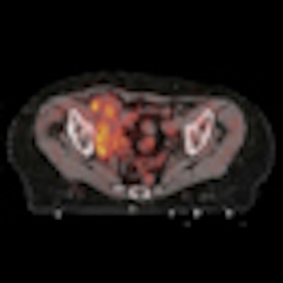
The results of a national survey of patient dose from whole-body PET/CT systems used in French nuclear medicine departments in 2011 have shown wide variations exist between centers, including fourfold differences in volume CT dose index (CTDIvol) levels.
"Practice seems to be different from one nuclear medicine department to another," noted lead author Cécile Etard, a researcher from l'Institut de Radioprotection et de Sûreté Nucléaire (IRSN) in Fontenay-aux-Roses, France. "In terms of the FDG activity and the DLP [dose-length product], the average effective dose related to a whole-body PET/CT examination in France can be assessed to be 14.3 mSv (applying International Commission on Radiological Protection [ICRP] 60 tissue weighting factors), with 8.6 mSv due to the whole-body CT scanner and 5.7 mSv due to the FDG-PET examination."
Current diagnostic reference levels (DRLs) for the diagnostic trunk CT are too high for CT combined with PET, although the examination is only performed in France for attenuation correction and localization, and not for diagnostic purposes, she wrote in an online article published by Radiation Protection Dosimetry on 14 May. She proposed DRLs equal to 8 mGy (CTDIvol) and 750 mGy cm (DLP) for whole-body PET/CT. The average effective dose related to whole-body PET/CT examination in France has been assessed to ~14 mSv.
Characteristics of the 56 PET/CT units included in the survey: Manufacturer, type of scintillator, commissioning year, and time-of-flight (TOF) technology when present
|
||||||||||||||||||||||||||||||
The survey was conducted in July 2011 by emailing one questionnaire to all members of the French Society of Nuclear Medicine (SFMN). Only departments equipped with a PET/CT unit were asked to respond. The survey was dedicated to FDG examinations combined with whole-body scanning. The objectives were to provide accurate data on the patient dose related to PET/CT, focusing on CT, and to propose a national DRL for a CT examination combined with PET examination.
The first part of the questionnaire related to the equipment: manufacturer, model, and commissioning date. The second part dealt with dosimetric data. Medical staff had to register the age, weight, injected FDG activity, CTDIvol, and DLP for 20 PET/CT examinations performed on patients weighing from 50 to 100 kg. These two dosimetric quantities are available on every CT model, and the periodic mandatory quality control of CT scanners includes their control. In the case of whole-body CT, they are related to a 32-cm diameter phantom, according to Etard and colleagues.
The results, in terms of the FDG activity and the specific activity: CTDIvol and DLP were analyzed and compared with the current DRLs for FDG and diagnostic CT, and with the European guidelines for tumor PET imaging. The average effective dose per PET/CT examination was estimated using the ICRP conversion factor for the PET contribution. The CT contribution to the average effective dose was estimated using CTexpo V2.0.1 software for different types of CT scanners in the survey, including GE Healthcare's Brightspeed 16, Philips Healthcare's Brilliance 16 and Siemens Healthcare's Sensation 16.
In 2011, 90 PET units were operating in France in 198 nuclear medicine departments. The study involved 56 PET/CT units operating in 55 nuclear medicine departments. The response rate for the survey was 62%, and the study included about 1,000 examinations. Seven departments sent data related only to CT examinations. The three manufacturers were almost equally represented in the sample, the authors stated.
The table shows the geographical distribution and the characteristics of the PET/CT units in the survey. Time-of-flight (TOF) technology, available in most newer models, was used in 15 of the 56 systems. Except for one unit, the CT scanners were multislice machines (two to 64 slices), and two of them used an iterative reconstruction algorithm.
The average specific injected FDG activity was equal to 4.3 MBq kg-1, in agreement with European recommendations. The TOF technology led to a reduction in the specific activity to 3.5 MBq kg-1. Data related to the FDG activity were available for 49 of the PET/CT units. The average FDG specific activity was equal to 4.3 MBq kg-1, in line with the European recommendations (2.5-5 MBq kg-1).
"For six departments, the specific activity was significantly higher than this recommendation. For most of the nuclear medicine departments, the specific activity was almost constant and the injected FDG activity was adapted to the patient weight," Etard explained.
As for the distribution of the injected FDG activity for all examinations in the survey, the average value was equal to 301 MBq, less than the current national DRL equal to 350 MBq. Using the ICRP conversion factor for FDG, this average activity corresponds to an average effective dose equal to 5.7 mSv, she noted.
The average value and the 75th percentile of the CT dose index distribution were equal to 6.6 mGy and 7.7 mGy. The 75th percentile was much lower than the national DRL for the diagnostic trunk CT (20 mGy).
For the 1,000 examinations in the survey, the average value and the 75th percentile of the DLP distribution were 628 mGy cm and 762 mGy cm, respectively. As the exposed length is often longer than the trunk, this value cannot be compared with the DRL for the diagnostic trunk CT, the authors wrote. The effective dose associated with a DLP equal to 628 mGy cm for a whole-body CT (from neck to thighs) was estimated using CTexpo V2.0.1 software to 8.6 mSv.
For CTDIvol distribution, no major difference was identified among the three manufacturers or with the age of the CT unit, but the analysis of the average CTDIvol showed a fourfold factor among the 56 CT units included in the survey (from 3.4 mGy to 13.7 mGy). This may be explained partly by the routine protocol (high voltage, charge per slice, slice width, pitch). Lower voltage and charge per slice, as well as a higher pitch value, should help reduce the CTDIvol, and the associated decrease in image quality may be acceptable on CT, the authors wrote.
The values of 8 mGy and 750 mGy cm could be proposed as national DRLs in terms of CTDIvol and DLP, respectively, for a "whole-body" CT combined with PET examination, according to the "75th percentile methodology."
The relatively high level of participation and the homogeneous geographic repartition of the nuclear medicine departments included should help maximize the usefulness of this study, the authors stated. Also, there is an ongoing study that will bring elements to understand the large dispersion in patient doses among the nuclear medicine departments, especially regarding CT contribution. A national protocol established at the end of this study and recommended at the national level by bodies such as SFMN would facilitate the homogenization of the clinic practice and dose optimization, they concluded.