A digitally reconstructed radiograph computer algorithm has been produced -- and validated -- that adequately simulates anatomical and system noise. The image evaluators grade simulated chest x-rays at different exposure parameters to optimize radiographic technique for clinical computed radiography (CR) without repeat patient exposures.
Patient anatomy is the limiting factor in detecting lesions in the chest. Therefore, images used to optimize any digital x-ray system for chest radiography must contain clinically realistic features. Typically, CR chest optimization has used physical phantoms, but these do not necessarily contain all the anatomical features required. A possible solution is to use clinical images to obtain the "highest validity for optimization," using "a method to simulate lower exposures by adding frequency-dependent noise to an original image," according to the authors of a new study in the British Journal of Radiology (BJR, October 2011, Vol. 84:1006, pp. 890-902).
However, study author Craig Moore, a physicist and radiation protection adviser at Queen's Centre for Oncology and Haematology, Castle Hill Hospital in Cottingham, U.K., and colleagues point out this methodology only works for a given x-ray beam quality, so full optimization would still necessitate repeat exposures of the same patient, thus increasing the stochastic risk of inducing cancers. Moore and colleagues developed and validated a digitally reconstructed radiograph (DRR): a computer simulation of a conventional 2D x-ray image created from CT data using realistic anatomical and frequency dependent system noise.
"In our new model, a raycasting method of DRR generation was used, as this method has been proven to produce superior quality in the resultant images than those produced by other methods such as splatting," the researchers wrote in their study.
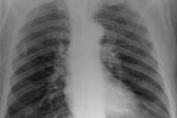
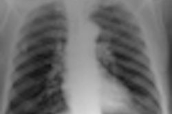
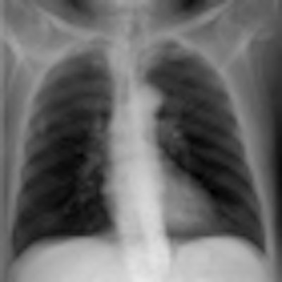
 Top: Digitally reconstructed radiograph (DRR) of a chest x-ray reconstructed with a tube potential of 50 kVp. Below: DRR of a chest x-ray reconstructed with a tube potential of 150 kVp. Bottom: DRR of a chest x-ray reconstructed with an antiscatter grid. There is increased detail in the spine and diaphragm region. All images courtesy of Craig Moore.

The x-ray spectra are generated by the Institute of Physics and Engineering in Medicine Report 78 software. Energy absorbed in the CR storage phosphor, scatter, and frequency-dependent noise are modeled according to physical characteristics of the CR system.
The model was validated with a phantom and patients; it provided predictions of signal-to-noise ratios, tissue-to-rib ratios, and pixel value histograms that lie within the range of values measured with patients and the phantom. The maximum difference in measured signal-to-noise ratios to that calculated was 10%. Tissue-to-rib ratio values differed by a maximum of 1.3%.
"Our DRR calculator scheme provides a realistic model of the Agfa HealthCare CR digital imaging system for chest radiography (of average size males)," the authors stated.
They found anatomical noise was adequately simulated using human chest CT datasets. Scatter and system noise was successfully added post-DRR calculation, which resulted in signal-to-noise ratios, tissue-to-rib ratios, and pixel value histograms that are in good agreement with those measured in CR-acquired images.
"Although DRR resolution is not as good as real CR images, four independent expert image evaluators believe DRR images adequately simulate real radiographs and provide realistic anatomical features. Therefore, this computer model provides us with a tool that can be used by radiologists to grade quality for images derived with different x-ray system parameters, without the need to perform repeat exposures on patients," they wrote.
The simulator can be adapted to include other digital detector technologies if x-ray photon absorption, noise, and scatter properties of the system in question are measured and incorporated into the simulation, they added.
In an interview with AuntMinnieEurope.com, Moore said the algorithm is exciting because of the following:
- Patients do not need any repeat exposures.
- Numerous different radiographic techniques can be simulated.
- The algorithm can be used for other types of digital detectors, not just CR.
"We have already used computer-generated images in our Hospital Trust to optimize chest CR imaging," he noted.
Simulated images reconstructed at various x-ray exposure parameters and techniques were scored by experienced image evaluators; optimum tube potential, scatter rejection technique, and receptor doses for clinical CR chest radiography have been derived, he added.
"At the outset of this work, CR chest exposure factors across the Hull and East Yorkshire Hospitals NHS Trust (HEY) were not standardized, and therefore not optimized," he said. "This work concluded with recommendations to the HEY radiology department for optimum exposure factors and technique for chest radiography. These were implemented across the Trust as a result of this work."
In their study, the researchers stated that future work will include adding pathology to the CT data to simulate lesions in the resulting DRR, modeling antiscatter grids and air-gap techniques for scatter reduction investigations, and simulating large/obese patients.
Results of the optimization study have been accepted for publication in the BJR and should be out in 2012, according to Moore.