The revolution in imaging driven by digital radiography (DR) detectors is far from over. Faster access to images, file sharing with other physicians, and advances such as computer-aided diagnosis (CAD) have already convinced the medical community to convert from analog to digital. Yet the struggle for improved image quality continues, according to Dr. Thomas Vogl, who leads a research and development group at the Johann Wolfgang Goethe University Hospital in Frankfurt am Main in Germany.
Manufacturers of DR detectors, including Fujifilm Medical Systems USA, Agfa HealthCare, Carestream Health, Hologic, Toshiba Medical Systems, Canon U.S.A., GE Healthcare, Philips Healthcare, Siemens Healthcare, Imix, Swissray International, Imaging Dynamics, and Delft Diagnostic Imaging, the parent company of PACS developer Rogan-Delft, turn to Vogl's team to refine their products and explore new technological themes for enhancing image acquisition.
"We see direct detectors as a small revolution," he noted. "It is very spectacular and we are working hard on several programs to advance this revolution."
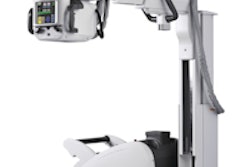
The multifunctional panels can be used in a number of clinical settings, including intensive care for bedside examinations, while portable DR detectors have created new applications in trauma, pediatric, and orthopedic settings.
The industry's first detector featuring wireless image transmission, the DRX-1 from Carestream, was introduced in 2009 at the University Hospital in Frankfurt and subjected to a series of studies by Vogl's group against conventional computed radiography (CR) systems for both image quality and workflow efficiency. As a result of the positive findings from the studies for superior image quality and dose efficiency, the DRX-1 detector is now used as part of routine protocol.

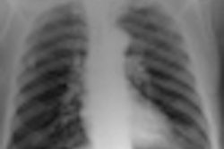
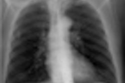
 Two images of a hand phantom obtained using different equipment: Fuji's CR system and DRX-1 from Carestream. All images courtesy of Dr. Thomas Lehnert.
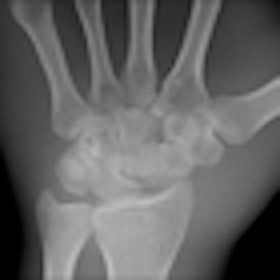
 A third image of a hand phantom obtained using DR9500 from Carestream.
Identical in size to a standard CR cassette, the detector can be inserted into an existing wall stand or table Bucky and used in combination with existing x-ray generators from a number of original equipment manufacturers, presenting an attractive option for modest-budget facilities. The flexibility of the portable detector provides great advantages in time-critical environments where an x-ray image can now be captured and viewed in less than six seconds, he explained.
Further advances in image quality are being explored in three approaches currently underway with the group at Goethe University to enhance the physical properties of the various detector systems. The most advanced program is exploring the integration of a pixilated superstructure of optically isolated cells into the cesium iodide scintillator to better suppress fluorescent light scatter. The group is only at a mid-way point in its experiments, but the data look quite promising, Vogl reported.
"We have a group of young people working on the program who are very enthusiastic, and actually we are having a lot of fun," he said. "You can expect to see upcoming articles in professional journals."
A second approach to enhancing indirect-conversion detectors is to pack powdered phosphors into a pixilated superstructure of cellular cavities separated by walls made of a reflective material to prevent cross-talk between the pixels and minimizing blurring. The group has performed 30% of the experiments designed to advance this investigation.
"There is a potential here, but is not yet as promising as the first approach, which is the priority among our programs," he said.
Further ahead is a program for enhancing image acquisition in direct conversion detectors by replacing the amorphous selenium used as the semiconductor material with an alternative photoconductor for greater x-ray absorption efficiency across the radiographic energy spectrum, as well as a greater yield of signal electrons.
Polycrystalline materials being evaluated as candidates for this role include lead iodide, mercury iodide, lead oxide, and cadmium-zinc-telluride.
"This program is in a very early stage and remains largely a theoretical question," Vogl said. "We have conducted some experiments using different materials, but I would not even call it an investigation so much as a preliminary look at how to investigate this question."