Medical physics experts may now use the image quality metrics described in a British Journal of Radiology study in quality assurance (QA) programs and optimization studies with a degree of confidence -- they reflect the clinical image quality in chest computed radiography (CR) images acquired without an antiscatter grid.
The results of the study support the value of using contrast-to-noise ratio (CNR) and effective dose efficiency (eDE) in the evaluation of quality in clinical thoracic radiography.
Chest radiography is one of the most frequently performed diagnostic radiographic examinations in the U.K. according to a Health Protection Agency report, so optimization of radiation dose and image quality in chest radiography is an important research area, especially in the era of digital imaging, according to a team led by Craig Moore, PhD, a medical physicist at the radiation physics department at Queen's Centre for Oncology and Hematology at Castle Hill Hospital in Hull (BJR, 17 May 2013).
"Many investigators have shown that it is not the system (including quantum) noise that is the limiting factor in chest radiography, but rather the projected patient anatomy or 'anatomical noise.' It therefore follows that any images used to optimize a digital radiographic system for chest radiography must contain clinically realistic anatomical features and noise," Moore and colleagues wrote.
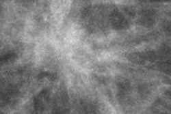
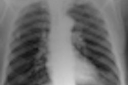
 Above and below, simulated chest radiographs of an average sized patient reconstructed at tube voltages of 50 kVp (above), and 125 kVp (below). Images courtesy of Craig Moore, PhD.
The metrics usually used to measure image quality have been found to be unique to individual detectors and are not measured under typical clinical conditions, so an alternative metric has been proposed: the effective detective quantum efficiency (eDQE). It provides a measure of the signal-to-noise transfer characteristics of a digital imaging system measured under clinical conditions, using a phantom designed for a specific examination type.
However, eDQE doesn't take into account the radiation risk to the patient, so the concept of eDE also was proposed, which is the effective noise equivalent quanta (eNEQ) normalized to the effective dose.
Moore and colleagues used the results of their previous optimization study of computer-simulated posteroanterior chest images (BJR, September 2012, Vol. 85:1017, pp. e630-e639) to investigate the images' correlation with the physical image quality metrics, eDE and CNR, across the diagnostic energy range (50-125 kV).
"This work examines the correlation between perceived clinical image quality (i.e., how good the images look as perceived by radiologists) to that which something medical physicists such as myself can actually measure objectively (physical image quality)," Moore said in an interview with AuntMinnieEurope.com.
Methods and results
To measure clinical image quality, the researchers used results of their previously published study in which four experienced image evaluators graded computer-simulated posteroanterior chest images using a visual grading analysis scoring scheme. For physical image quality, eDE was measured in a uniform chest phantom. Correlation between the two was determined using Pearson's correlation coefficient or R.
The researchers found statistically significant correlations between the visual scores that represented clinical image quality and the CNR representing physical image quality. For CNR, R = 0.87 (p < 0.033), while for eDE, R = 0.77 (p < 0.008).
"Medical physicists may use the physical image quality metrics described here in quality assurance programs and optimization studies with a degree of confidence that they reflect clinical image quality in chest CR images," Moore said. "This is potentially an important new step because it links what we can measure with phantoms with clinical image quality."
On the basis of this research, it may be possible to establish baseline physical metrics for chest radiography that correspond to the minimum clinical image quality required, he added.
"The availability of such baseline measurements will be extremely useful for medical physicists within a routine quality assurance program and for assessing how the introduction of new radiographic techniques (such as new exposure factors) impacts clinical image quality," Moore said.
As well as the link between clinical and physical image quality, the results of this paper also demonstrate that chest CR can be carried out with tube voltages much lower than those used traditionally, given that both clinical and physical image quality improve as tube voltage is decreased, he added.
It should be noted that the findings in this paper are specific to CR receptor technology from Agfa HealthCare. However, "given that most powder-based CR phosphors are based on the barium fluorohalide family, it is likely that the conclusions of this work are transferable to other manufacturers, but further studies would be worthwhile," Moore and colleagues wrote in the study.