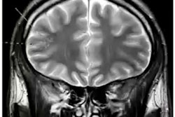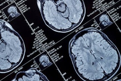
Japanese researchers have published a database that includes images from functional MRI (fMRI) cases of multiple clinical disorders, according to a paper published on 30 August in Scientific Data.
They hope the database will help further standardize artificial intelligence (AI) technology for use in medical imaging, specifically MRI, wrote a team led by Saori Tanaka, PhD, of Advanced Telecommunications Research Institutes International in Kyoto, Japan.
"In medicine, progressively, AI technology is being used to analyze medical images, such as functional magnetic resonance images (fMRI), to help diagnose diseases ... and reproducibility is essential for practical application of this technology," the group noted.
Tanaka and colleagues assembled a database that consists of 2,414 samples from both patients and healthy controls of resting-state fMRI, structural MRI, and demographic information for a variety of psychiatric and neurological diseases, including autism spectrum disorders, major depression, bipolar disorder, schizophrenia, obsessive-compulsive disorder, chronic pain, and stroke. The data were acquired from 14 sites.
"Researchers will have global access to these data, and the speed of research may be dramatically increased through development of more accurate diagnostic markers and more advanced harmonization methods," Tanaka's group concluded.
The data are available on Synapse with the ID code syn22317076.




![A normal mammogram confirmed by three-year radiologic follow-up illustrates reader-marked regions of interest (ROIs) during (A) unaided (round 1) and (B) artificial intelligence (AI)–assisted (round 2) reading. Each colored dot represents an ROI for recall by a human reader. Readers could mark more than one ROI per case, represented by multiple dots of the same color. During AI-assisted reading, the AI system displayed three visible prompts: two with suspicion of malignancy scores of 35% (left mediolateral oblique [L MLO] and craniocaudal [L CC]) and one with a suspicion of malignancy score of 10% (right craniocaudal [R CC]), shown as polygonal overlays. Without AI, six of 10 readers (60%) marked a false-positive ROI. With AI assistance, this fell to two of 10 (20%). R MLO = right mediolateral oblique.](https://img.auntminnieeurope.com/mindful/smg/workspaces/default/uploads/2026/07/2026-07-14-radiology-mammogram-ai-auto-bias.H0bYO8QlWs.jpg?auto=format%2Ccompress&fit=crop&h=100&q=70&w=100)


![A normal mammogram confirmed by three-year radiologic follow-up illustrates reader-marked regions of interest (ROIs) during (A) unaided (round 1) and (B) artificial intelligence (AI)–assisted (round 2) reading. Each colored dot represents an ROI for recall by a human reader. Readers could mark more than one ROI per case, represented by multiple dots of the same color. During AI-assisted reading, the AI system displayed three visible prompts: two with suspicion of malignancy scores of 35% (left mediolateral oblique [L MLO] and craniocaudal [L CC]) and one with a suspicion of malignancy score of 10% (right craniocaudal [R CC]), shown as polygonal overlays. Without AI, six of 10 readers (60%) marked a false-positive ROI. With AI assistance, this fell to two of 10 (20%). R MLO = right mediolateral oblique.](https://img.auntminnieeurope.com/mindful/smg/workspaces/default/uploads/2026/07/2026-07-14-radiology-mammogram-ai-auto-bias.H0bYO8QlWs.jpg?auto=format%2Ccompress&fit=crop&h=112&q=70&w=112)