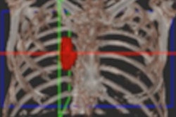
Advanced visualization and artificial intelligence (AI) software developer TeraRecon has introduced two new lung density analysis software applications to aid in the assessment and follow-up care of patients with COVID-19.
Lung Density Analysis II (LDA-II) enables TeraRecon's Intuition customers to upgrade their advanced visualization software to include a custom lung workflow. Making use of Intuition's lung segmentation, volumetric histogram analysis, and automation capabilities, LDA-II provides physicians with colorized densities and lung composition values that can be analyzed and quantified with little to no manual editing, the company said.
Current Intuition customers can access LDA-II via a remotely installed upgrade. The software is also included with a subscription to the firm's Intuition Titanium Suite.
The second new offering, Emergency Lung AI Suite, is designed to provide fast access to lung density analysis tools across the enterprise and to physicians working remotely, according to the vendor. A cloud-based platform, Emergency Lung AI Suite enables physicians to upload cases, anonymize patient data, process the LDA-II algorithm, and be notified of results.
No installation is required, and the software can be quickly integrated with diagnostic and point-of-care software applications. Emergency Lung AI Suite can also be extended to other clinical use cases within a health system's imaging and crisis readiness infrastructure, TeraRecon said.




![A normal mammogram confirmed by three-year radiologic follow-up illustrates reader-marked regions of interest (ROIs) during (A) unaided (round 1) and (B) artificial intelligence (AI)–assisted (round 2) reading. Each colored dot represents an ROI for recall by a human reader. Readers could mark more than one ROI per case, represented by multiple dots of the same color. During AI-assisted reading, the AI system displayed three visible prompts: two with suspicion of malignancy scores of 35% (left mediolateral oblique [L MLO] and craniocaudal [L CC]) and one with a suspicion of malignancy score of 10% (right craniocaudal [R CC]), shown as polygonal overlays. Without AI, six of 10 readers (60%) marked a false-positive ROI. With AI assistance, this fell to two of 10 (20%). R MLO = right mediolateral oblique.](https://img.auntminnieeurope.com/mindful/smg/workspaces/default/uploads/2026/07/2026-07-14-radiology-mammogram-ai-auto-bias.H0bYO8QlWs.jpg?auto=format%2Ccompress&fit=crop&h=100&q=70&w=100)


![A normal mammogram confirmed by three-year radiologic follow-up illustrates reader-marked regions of interest (ROIs) during (A) unaided (round 1) and (B) artificial intelligence (AI)–assisted (round 2) reading. Each colored dot represents an ROI for recall by a human reader. Readers could mark more than one ROI per case, represented by multiple dots of the same color. During AI-assisted reading, the AI system displayed three visible prompts: two with suspicion of malignancy scores of 35% (left mediolateral oblique [L MLO] and craniocaudal [L CC]) and one with a suspicion of malignancy score of 10% (right craniocaudal [R CC]), shown as polygonal overlays. Without AI, six of 10 readers (60%) marked a false-positive ROI. With AI assistance, this fell to two of 10 (20%). R MLO = right mediolateral oblique.](https://img.auntminnieeurope.com/mindful/smg/workspaces/default/uploads/2026/07/2026-07-14-radiology-mammogram-ai-auto-bias.H0bYO8QlWs.jpg?auto=format%2Ccompress&fit=crop&h=112&q=70&w=112)