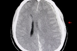
A set of artificial intelligence (AI) algorithms can prescreen head CT scans for urgent findings such as intracranial hemorrhage and cranial fracture, enabling triage of these cases for priority review by radiologists, according to research published online on October 11 in Lancet.
Researchers led by Sasank Chilamkurthy of AI software developer Qure.ai found that deep-learning algorithms could be highly accurate for separately detecting up to nine critical findings.
"The strong performance of deep-learning algorithms suggests that they could be a helpful adjunct for identification of acute head CT findings in a trauma setting, providing a lower performance bound for quality and consistency of radiological interpretation," the authors wrote.
Noncontrast head CT studies are commonly used as a first-line diagnostic method in emergency rooms for patients with a head injury or with symptoms suggesting a stroke or an increase in intracranial pressure. Intracranial hemorrhages, raised intracranial pressure, and cranial fractures are the most critical and time-sensitive abnormalities that can be readily detected on these studies, according to the researchers.
"Although these abnormalities are found on only a small proportion of CT scans, streamlining the head CT scan interpretation workflow by automating the initial triage process has the potential to substantially decrease time to diagnosis and expedite treatment, which might in turn decrease morbidity and mortality consequent to stroke and head injury," the authors wrote. "An automated head CT scan triage system might also be valuable for queue management in a busy trauma care setting, or could facilitate decision-making in remote locations without availability of an immediate radiologist."
The researchers developed deep-learning algorithms to separately detect up to nine critical findings: intracranial hemorrhage and its five types (intraparenchymal, intraventricular, subdural, extradural, and subarachnoid); calvarial (cranial vault) fractures; midline shift; and mass effect.
They retrospectively gathered 313,318 anonymous head CT scans and their associated radiology reports from around 20 centers in India to train and perform initial validation of the algorithms. The algorithms were then tested on an additional dataset of 491 scans gathered from six other hospitals. Consensus diagnosis of three independent radiologists served as the gold standard for the test dataset.
| Performance of AI algorithms on test set of head CT scans | |
| Condition | Area under the curve |
| Intracranial hemorrhage (ICH) | 0.94 |
| Intraparenchymal ICH | 0.95 |
| Intraventricular ICH | 0.93 |
| Subdural ICH | 0.95 |
| Extradural ICH | 0.97 |
| Subarachnoid ICH | 0.96 |
| Calvarial fractures | 0.96 |
| Midline shift | 0.97 |
| Mass effect | 0.92 |
"Our results show that deep-learning algorithms can accurately identify head CT scan abnormalities requiring urgent attention, opening up the possibility to use these algorithms to automate the triage process," the authors wrote.
The researchers acknowledged, though, that it's possible that overreliance on such a triage approach might lead to automation bias in radiologists, resulting in false-negative scans being overlooked.
"A prospective clinical trial is necessary to determine the safety and efficacy of such a triage and if it ultimately improves patient care and outcomes," they wrote.