
Researchers from the U.K. and France collected prestigious prizes at the European Society for Magnetic Resonance in Medicine and Biology (ESMRMB) meeting in Toulouse, France, for their investigations into resting state functional MRI.
A group from King's College London (KCL) proposed a framework for motion correction in resting state functional MRI (rsfMRI) examinations in the moving fetus, and they received the Magna cum Laude award for their efforts when the ESMRMB congress ended on Saturday.
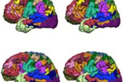
Resting state networks (RSNs) are coherent and spatially well located fluctuations of the blood oxygen level dependent (BOLD) signal, and they appear whenever the brain undergoes a resting state condition, according to lead author Giulio Ferrazzi, a research student in the biomedical engineering department, and his colleagues. Ferrazzi's lead supervisor -- Jo Hajnal, PhD, a professor and the chair of imaging sciences at KCL -- was a co-author of the award-winning e-poster.
"RSNs have been extensively mapped out in adults, where the amount of exhibited motion is relatively small," they explained. "The presence of RSNs in infants and preterm babies has also been proved, suggesting that this phenomenon starts to affect the functional development of the human brain from the first stages of life."
 The organizers of the ESMRMB congress chose the bell tower of the St. Sernin Basilica in Toulouse, France, as their main image. The church is located on the site of a previous basilica from the fourth century, which contained the body of Saint Saturnin or Sernin, the first bishop of Toulouse. Most of the current building was constructed in the Romanesque style between about 1080 and 1120, and it became a UNESCO World Heritage Site in 1998.
Motion corrupts slow imaging techniques such as MRI, rendering data acquisition and analysis challenging, and in most previous studies of RSNs in fetuses, as much as half (or even more) of the data were excluded because of motion. To combat this problem, the authors developed and tested a processing framework for fetal rsfMRI, capable of correcting gross motion. The method comprises slice to volume registration and target volume estimation, and they devised a simple way to cope with nonuniform temporal sampling of fetal fMRI data.
The team scanned eight fetuses (mean gestation of 30, std ± 5.96 weeks) on a Philips Healthcare Achieva 1.5-tesla system with a 32-channel receiver coil using single-shot echo-planar imaging (EPI) (TR = 4,000, TE = 50, 30 slices x 120 time points at 2.5 x 2.5 x 5 mm3). The processing framework was divided into two different parts. The first part corrects for motion, and the second for nonuniform temporal sampling of fetal fMRI data.
EPI represents one of the fastest sequences available in MRI, Ferrazzi and colleagues noted. In the single-shot mode, the acquisition of a single slice takes less than 100 msec, and intraslice motion artifacts can be effectively neglected. Due to the large amount of time that is usually required during acquisition, interslice motion is still present. Because the head of the fetus does not deform during acquisition, motion is also rigid.
The correction algorithm assumes rigid motion to be happening between slices, and aims to calculate the roto-translation matrices able to map data entries from the coordinate system of the scanner into the correct anatomical position, the authors wrote.
As a first step in the processing pipeline, single stacks are registered altogether and a reference image is formed by averaging the entire dataset. Data are then divided into temporally contiguous blocks, containing fewer slices. Rigid body registration onto the previous reference is then performed, and the reference image is updated. This process is repeated, with progressively reduced temporal groupings, stopping when each block contains one slice. The last iteration of the algorithm corresponds to slice to volume registration step. Each slice is then projected into a high-resolution anatomical space using the transformations so determined, to create fully motion-corrected fetal fMRI datasets, they stated.
In the time correction framework, nonuniform temporal sampling of fetal fMRI data will appear as a natural consequence of motion, where slices acquired in the coordinate system of the MRI unit are effectively sampling the fetal brain at different anatomical positions, Ferrazzi and colleagues said. To correct for this, pixels entries were mapped at the time scale of TS = TR/number of slices, data were then shuffled according to the slice time ordering, and the time series recovered back at the temporal resolution of TR using sinc interpolation.
"This study demonstrates that slice to volume registration with slice timing correction methods can enable group independent component analysis (ICA)-based analyses of fetuses with no data loss," they concluded. "Although as yet simple, the current procedure includes core requirements for motion and time corrections, creating foundations for a robust fetal fMRI approach."
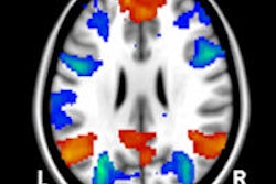
In another prize-winning e-poster presented at ESMRMB, French authors showed that resting-state fMRI studies reveal a profound reorganization in the brains of comatose patients.
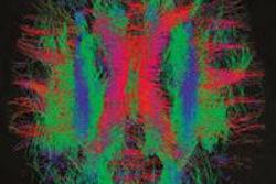
Sophie Achard, PhD, a research fellow in the Centre National de la Recherche Scientifique, GIPSA-lab in Grenoble, France, and her colleagues studied the graph representing individual brain connectivity at rest in a group of comatose patients and of healthy controls. They received a certificate of merit from the ESMRMB judging panel.
"Interestingly, we found that the graph's global topology in patients remain preserved," they noted. "To further study possible reorganization of nodes' connections, we introduced a new metric that we called the graph reorganization index, an index that is sensitive to the local variations of connectivity. Using this index, we could observe that hubs of the graphs in healthy controls become nonhubs in patients, and that nonhubs in healthy controls become hubs in patients. We can thus hypothesize that consciousness might be supported by the hubs' locations within the brain."