
Fusion of single photon emission CT (SPECT) and MR images, along with sequential gamma images, can improve the assessment of segmental gastrointestinal (GI) transit time by facilitating accurate delineation of the tracer anatomically, researchers from Malaysia and the U.K. have found.
"Comparing SPECT-MR post-fusion combines both morphologic and functional data, providing a more accurate localization of the site of problem in GI motility examinations," noted lead author Chai Hong Yeong, MSc, a medical physicist at the department of biomedical imaging, University of Malaya, Kuala Lumpur, Malaysia, in a prize-winning e-poster at last month's European Congress of Radiology (ECR).
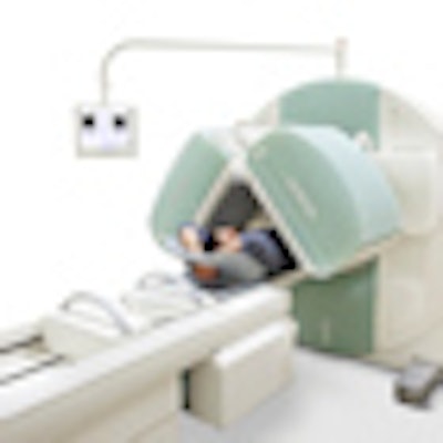
 When fused with MRI, images from a SPECT system like this can improve the assessment of segmental gastrointestinal transit time by facilitating accurate delineation of tracer anatomically. Image courtesy of Siemens Healthcare.
"The imaging procedures can be done using two separate modalities within a reasonable time frame, and the images are fused during postprocessing. At least four external body markers are needed to provide accurate landmark registration of two volumetric images," she stated.
The researchers' aimed to demonstrate the feasibility of SPECT and MR image fusion for improving the diagnosis of regional abnormalities in whole GI transit. For each enteric-coated Samarium-153 (Sm-153) radiolabelled capsule, 50 mg of Sm-152Cl3-6H2O was weighed and dissolved in 1 ml of distilled water, and 100 mg of Amberlite IR-120 (H+) ion-exchange resin was added to the solution and mixed thoroughly.
After the mixture was dried in a laboratory oven for 12 hours at a temperature of 70o C, the dried resin beads were put into a medical-grade size one empty hard gelatin capsule. The gelatin capsule was designed to remain intact within the stomach but to disintegrate when it reached the small intestine. Therefore, the capsule was enteric-coated with a biocompatible polymer solution containing 13% Eudragit L100.
The capsules were then sent for neutron activation using a 250 kW open pool-type research reactor (Triga Mark II, General Atomics). The capsules were irradiated in a neutron flux of 1 x 1013 cm-2s-1 for 100 s to achieve nominal radioactivity of 5 MBq after 66 hours of neutron activation, which was the intended diagnostic reference level (administered activity intended for clinical study), according to the authors.
They recruited 10 healthy volunteers (six females, four males, aged 33 ± 13) with no history of GI problems. Four external body markers containing 0.37 ± 0.12 MBq radioactive Sm-153 and 0.18 mgml-1 Gd-DTPA were placed on the left and right lower costal margins and iliac crests of each volunteer. Using a dual-detector SPECT system, one minute anterior and posterior scintigraphic images were acquired hourly for nine hours, followed by a final volumetric SPECT acquired with 64 angular steps over 3600 at 15 seconds per view in 64 x 64 matrix size. Subsequently, a volumetric T1-weighted MR image (TR 4.0 ms; TE 2.0 ms; SL 2.0 mm; 156 slices) was acquired using a 1.5-tesla MR system.
The researchers used MIPAV (Medical Image Processing, Analysis and Visualization) software to process and fuse the volumetric images from SPECT and MRI. The SPECT images were first reconstructed to the coronal plane to be aligned with the coronal volumetric image from MRI. The SPECT coronal images were then registered to the MR images using least squares method based on the four external body markers. The fused 3D image was then saved into a multimodalities DICOM format. Total whole body effective dose per subject was 0.88 ± 0.16 mSv.
There is increasing interest in measurement of whole gut motility for the examination of regional delayed GI transit, Yeong explained.
Gamma scintigraphy can be performed in conjunction with the oral administration of a nonabsorbable radiotracer. Indium-111 (In-111) is a commonly used radiotracer for the study of whole gut motility because of its long physical half-life of 67.3 hours, but it is not always available and shipping costs can be high. Also, the radionuclide must be incorporated into a suitable nonabsorbable formulation prior to use. This is often performed manually within the hospital radiopharmacy units and may result in personal radiation exposure to the individuals carrying out the procedure, she noted.
Yeong received a certificate of merit for her ECR 2012 exhibit. One of her co-authors was Alan Perkins, PhD, professor of medical physics at Queen's Medical Centre, Nottingham, who is president elect of the British Nuclear Medicine Society. He is currently involved in establishment of cyclotron and PET imaging facilities for Nottingham hospitals.
References
- Yeong CH, Abdullah BJJ, Ng KH, et al. Production and first use of 153SmCl3-ion exchange resin capsule formulation for assessing gastrointestinal motility. Appl Radiat Isot. 2012;70(3):450-455.
- Yeong CH, Abdullah BJJ, Ng KH, et al. Neutron activated 153Sm-ion-exchange resin as a tracer for gastrointestinal scintigraphy. Nuc Med Comm. 2011;32(12):1256-1260.
- Yeong CH, Blackshaw PE, Ng KH, et al. Reproducibility of neutron activated Sm-153 oral dose formulations intended for human administration. Appl Radiat Isot. 2011;69(9):1181-1184.